Sunday, March 22, 2015

Mitral Regurgitation


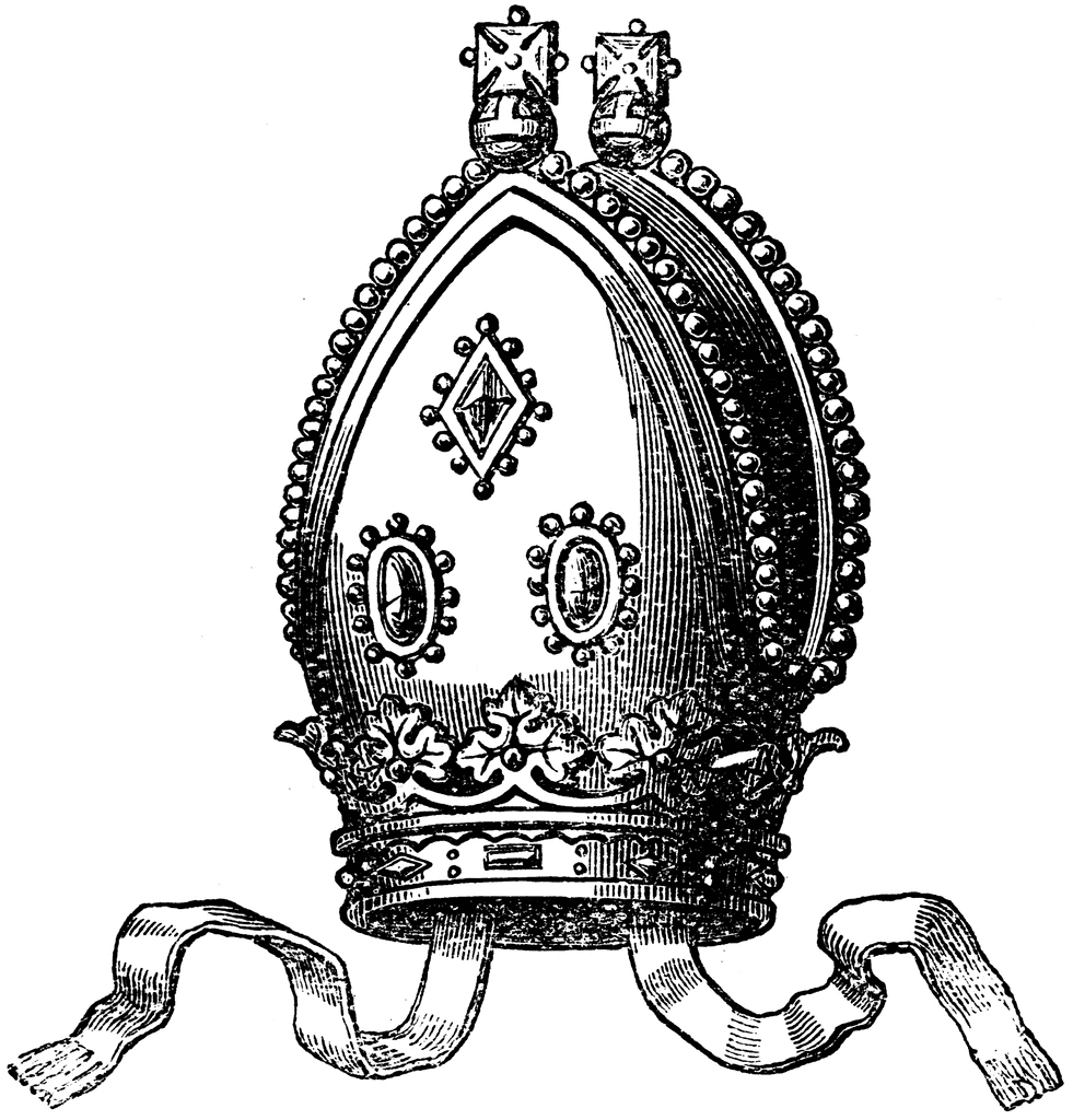
Mitral regurgitation is a common valvular problem seen on the medical wards. The valve is named after a mitre, the headgear worn by bishops, due to its resemblance. Here's a quick review of the management of this valvular problem.

Causes
- Primary causes: MVP, rheumatic disease, IE, trauma, myxomatous changes.
- Secondary: Ischemia, HCM, LV dilation, other valvular lesions (AS, AI)

Symptoms
- Most are asymptomatic for years, even with severe disease.
- Common presentation is intermittent atrial fibrillation.
- Other symptoms: shortness of breath, fatigue, pulmonary edema, and peripheral edema.
- 5 year survival: 80% if asymptomatic, 45% if symptoms.

Physical Examination
Holosystolic, blowing murmur. If severe, thrill and S3 are palpable.
- Arterial pulse is brisk
- Apical impulse is hyperdynamic


Investigations
Echo
  • Mitral annular calcification (MAC) - Crescent shaped calcification, generally associated with aging; associated with MR
  • No calcification - Myxomatous mitral valve disease (MVP); associated with MR
  • Leaflet calcification and subvalvular thickening - Rheumatic mitral valve disease; associated with MS
- If the cause of MR is not obvious, then the patient should have TEE.
- Severe MR must also include LA enlargement or LVH
- Severity of LA enlargement may correlate with chronicity of MR.
- Severe MR: Regurgitant fraction of greater than 50%; ERO greater than 0.4.

Coronary Angiogram:
  • Assess hemodynamics and severity of MR when noninvasive tests are inconclusive or discordant and for definition of coronary anatomy when patients are being considered for an intervention. 

                                        
                                Blood prefers to go back into the LA than the systemic circulation due
                                to lower pressures, creating a state of reduced afterload for the LV.

Indications for Surgery

Chronic
  1. Severe, symptomatic, and EF greater than 30%/LVEDP less than 50 mm Hg
  2. Severe, asymptomatic, and EF 30-60%
  3. Normal EF AND atrial fibrillation or pulmonary hypertension
- Optimize medically before surgery as much as possible - afterload reduction is preferable
- Repair is preferred over replacement
- Treat at compensated phase and before decompensated stages of disease
- EF must be greater 30% and LVEDP less than 50 mm Hg for MVR to be successful 
- If EF more than 50% and symptomatic, then consider surgery.
- MVR for anyone with symptoms; if uncertain, then do exercise testing. 


Acute
  1. Symptomatic 
  2. Flail valve
Why a picture of Elizabeth Taylor? She had mitral regurgitation and had valve replacement surgery at the age of 77.

Monday, March 16, 2015

Crystal Arthropathy

Let's talk about a common problem seen on Internal Medicine - Crystal Arthropathy
                           
Gout - Negatively birefringement crystals
CPPD - Weakly positive rhomboid crystals

CPPD 
  • Most are idiopathic; inflammation due to calcium deposit in the joint
  • Remember 3 Hs 
    • Hemochromatosis
    • Hyperparathyroidism
    • Hypomagnasemia (Gittelmans, Bartters)
  • Some more H's - hypothyroidism, hypophosphatemia, hypomagnesemia
  • Hemochromatosis - 2nd + 3rd MCP/PIP
  • Suspect CPPD when the presentation sounds like OA in odd locations (eg MCP) with chrondrocalcinosis
  • Basic Calcium Phosphate (BCP) - Milwaukee shoulder - destructive, acute, older females, associated with prior trauma
  • Arthrocentesis:  WBC 2000 - 10000. PMNS greater than 50%.
  • Acute treatment: Joint aspiration/steroid injection is first line
    • NSAID is second line
    • Colchicine, steroids (systemic or intra-articular) also used
    • Prophylaxis with colchicine if more than 2 attacks/year
                 

GOUT

Secondary Causes
  • PRV, hemolysis, TLS
  • Diet - beer, red meat, seafood
  • Increased meat, seafood, alcohol
  • Male, obesity
  • HTN
  • CKD
  • Leukemia/Lymphoma
  • Drugs: Cyclosporine, salicylates, HCTZ
Non-Pharmacologic Treatment
  • Lose weight
  • Change diet - increase protein
  • lower alcohol
  • Remove diuretic, change to losartan
Pharmacologic Therapy
  • Start ASAP
  • Treat with NSAIDs: 
    • eg Naproxen 500 mg bid or indeomethacin 50 mg tid. 
    • Duration: until 1-2 days after symptoms resolve (usually 5-7 days). 
    • Avoid ASA - will make it worse.
  • If contra-indicated, treat with colchicine 1.5 mg q8H x3, then taper until resolution. Colchicine is initiated in acute attacks only.
    • Only start this if symptoms are less than 48 hours
    • Don't use IV colchicine.
  • If contra-indicated, treat with oral, intra-articular, or IV glucocorticoids. 
    • Use only steroids for CKD and ESRD.
    • Intra-articular injection for those with 1-2 affected joints who can't take above medications.
    • Prednisone 30-50 mg daily over 7-10 days.
    • Intravenous - if all the above is contra-indicated; 20 mg methylprednisone IV.
  • Prophylaxis with urate lowering therapy in these people. 
    • 2 or more attacks/year
    • Tophi
    • Kidney stones
    • Radiographic changes
    • Urinary uric acid greater than 1100 mg/day
  • Which agent, how much?
    • Don't start during acute attack
    • Even when stable, start with overlap with NSAID/colchicine. Preferably Colchicine - continue for 6 months after normal urate is achieved.
    • Titrate to urate level less than 357 umol
    • Renal dose adjustment
    • Start with allopurinol; if intolerant, move on to febuxostat
    • Don't use these two agents when the patient is also on azathioprine
      • In this case, use probenacid instead (causes stones - make sure to hydrate)
    • If very severe, treat with IV pegloticase Q2 weeks