Monday, November 26, 2012

SVC Syndrome


Today in noon report we talked about a patient with an anterior superior mediastinal mass. 

The differential for these can be remembered as the five Ts:
- thyroid mass
- thoracic aorta
- teratoma
- thymoma
- terrible lymphoma

Sometimes these masses can compress the Superior Vena Cava, causing SVC Syndrome. 

Clinical Features of SVC Syndrome:
- Facial and neck edema, plethora 
- Venous collaterals visible over chest
- Stridor, hoarseness, cough (edema causing pressure on larynx)
- Dysphagia (edema causing pressure on pharynx)
- Cerebral edema: headaches, confusion

Causes of SVC Syndrome: The first step is to distinguish thrombosis from extrinsic compression by a mass. This is usually done with a contrast CT scan. Thrombosis is usually a result of instrumentation (catheters, pacemaker wires) while compression is usually from malignancy (lung, lymphoma or metastatic).

Treatment of SVC Syndrome:
- glucocorticoids
- radiation- when a tissue diagnosis has been made
- chemotherapy in non-Hodgkin's lymphoma and small cell lung cancer
- stenting by interventional radiology
- removal of hardware and/or local thrombolysis for acute thrombosis followed by anticoagulation (not indicated if chronic)


Monday, November 19, 2012

Psoriatic Arthritis


Today in morning report we talked about the physical exam of a patient with psoriasis, focusing on evidence of psoriatic arthritis

Here are some tidbits that came up:

1) Psoratic Arthritis Presentation:
- 60–70% of cases, psoriasis precedes joint disease.
- 15–20% of cases, the two manifestations appear within 1 year of each other.
- 15–20% of cases, the arthritis precedes the onset of psoriasis

There are five classic patterns of presentation:
- Isolated DIP arthritis
- Asymmetric oligoarthritis
- Symmetric polyarthritis (similar to RA)
- Axial disease (sacroiliitis & spondylitis)
- Arthritis mutilans

Extra-articular changes:
- Nail changes are present in 90% of cases, there are six patterns: pitting, horizontal ridging, onycholysis, yellowish discoloration of the nail margins, dystrophic hyperkeratosis, and combinations of these findings
- Dactylitis ("sausage digit") in >30%
- Enthesitis/tenosynovitis
- Eye: conjunctivitis or uveitis


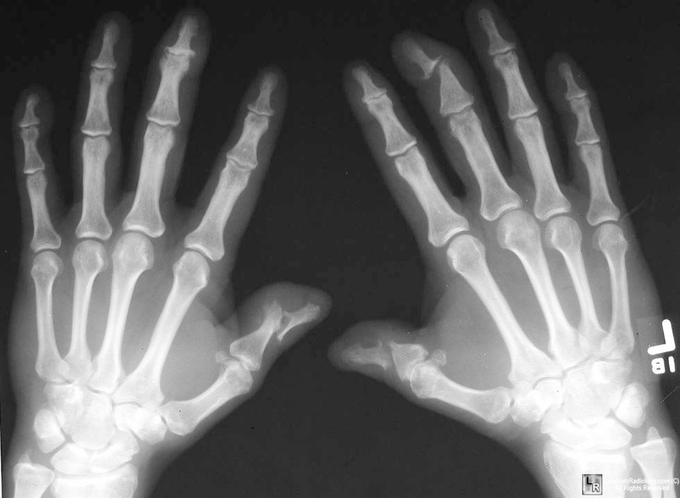
2) Findings on x-ray imaging of the hand in Psoriatic Arthritis
- DIP involvement, including the classic "pencil-in-cup" deformity (as seen above in top image)
- marginal erosions with adjacent bony proliferation ("whiskering")
- small-joint ankylosis (loss of space)
- osteolysis of phalangeal and metacarpal bone, with telescoping of digits (second image above)
- periostitis and proliferative new bone at sites of enthesitis.


3) Physical Examination for Axial Involvement: 
To Assess the sacroiliac joint: 
- palpation along SI joint
- FABERE test: flexion, abduction, external rotation and extension of affected leg (figure 4). Test is positive if tested leg cannot be lower parallel to other leg- this will occur with hip disease, iliopsoas spasm or SI joint disease.
- lateral compression of pelvis
- Gaenslen test (below):


To Assess mobility of lumbar spine:
- Modified Schober test: mark the point 10cm above and 5cm below the level of the dimple of Venus at the midline. Measure again with full forward flexion. This should increase to at least 20cm in flexion.
- standing back to a wall: distance from fingertip to floor on lateral flexion


4) BONUS! Diagnostic Criteria:

CASPAR criteria, a patient must have inflammatory articular disease (joint, spine, or entheseal) with 3 points from any of the following five categories:
  1. Evidence of current psoriasis, a personal history of psoriasis, or a family history of psoriasis
  2. Typical psoriatic nail dystrophy observed on current physical examination
  3. A negative test result for rheumatoid factor
  4. Either current dactylitisf or a history of dactylitis recorded by a rheumatologist
  5. Radiographic evidence of juxtaarticular new bone formation in the hand or foot

    That's all folks!


Wednesday, November 14, 2012

Myasthenia Gravis


Today in morning report we heard about a patient with dysarthria, dysphagia and ptosis. The diagnosis was Myasthenia Gravis.

Pathophysiology: 
- antibodies bind to the post-synaptic acetylcholine receptors
- this blocks acetylcholine binding, lead to crosslinking of receptors promoting internalization and degradation. 
- thus the number and availability of receptors are reduced
- thus end-plate potentials are insufficient to generate action potentials in a number of muscle fibers, causing weakness.

Presentation: 
- Up to 65% of patients initially have ocular symptoms of double vision and drooping of the eyelids.Less than 1/4 present with bulbar symptoms (dysarthria, dysphagia, altered voice). 
- Symptoms are typically better in the morning and after rest. 

Most Useful on history and physical:
- To rule in: “speech becoming unintelligible during prolonged speaking” (LR 4.5), peek sign (LR 30- illustrated in the third frame above), but their absence does not rule it out
- To confirm diagnosis or rule it out:  ice test (positive LR 24, negative LR 0.16), sleep test (positive LR 53, negative LR 0.01) or edrophonium (acetylcholinesterase inhibitor) test (positive LR 15, negative LR 0.11)

Confirming the diagnosis:
1. Acetylcholine receptor antibody test: most specific for MG. Sensitivity in generalized MG is 80%-96%,  but up to 50% of patients with purely ocular myasthenia are seronegative.

2. Single-fiber electromyography: highly sensitive for disorders of the neuromuscular junction but is not specific for myasthenia gravis.

Management:
- Rule out associated condition such as thymoma with CT chest and hyperthyrdoidism with TFTs. 
- Work up for other inflammatory conditions (ANA, RF)
- PFTs to assess for respiratory muscle weakness 

- Symptomatic treatment with pyridostigmine
- Indications for thymectomy: all patients with thymoma, some patients without thymoma (if generalised MG and under age 55 as 85% will have an improvement... however benefit takes years)
- Immunosuppression with glucocorticoids, azathioprine and others
- Avoid exacerbating drugs (including magnesium, beta-blockers, quinolones and macrolides)

Myasthenia Crisis:
- Myasthenic crisis is defined as an exacerbation of weakness sufficient to endanger life
- Close monitoring is key: Vital capacity (VC) and/or maximal inspiratory force (MIF) should be measured frequently, as often as every two hours. May need BiPAP or intubation if VC falling.
- Treat infectious triggers
- Immunomodulator: start prednisone or other immunosuppressive. With steroids, consider slow up-titration as high doses of steroids can exacerbate weakness.
-  IVIG 2g/kg over 5 days- improvement expected within a week
- Can also use Plasmapharesis but this is less well studied than IVIG
- Some advocate holding the anticholinesterase medication as "cholinergic crisis" can contribute to weakness

Tuesday, November 6, 2012

Myocarditis


Today in morning report we heard about a young woman with myocarditis.

Here were some key points:

1) Dangerous causes of chest pain in a young person:
Cardiac: 
- pericarditis
- myocarditis
- myocardial ischemia from drug use, thrombophilia, coronary artery dissection or premature atherosclerosis
- aortic dissection with condition predisposing to aortopathy such as Marfan's, Ehler's-Danlos, Ankylosing spondylitis or bicuspid aortic valve
Lung:
- pulmonary embolism
- pneumothorax
GI:
- Esophageal rupture

2) Causes of Myocarditis:
- Viral and post-viral: mostly Coxsackie, adenovirus, parvovirus and herpes viruses
- Bacterial: Borrelia burgdorferi and Ehrlichia species
- Parasites: Babesia, Trypanosoma cruzi
- Toxic: alcohol, radiation, chemicals (hydrocarbons and arsenic), and drugs, including doxorubicin
- Hypersensitivity: sulphonamides and penicillins
- Idiopathic: giant-cell myocarditis (autoimmune) and cardiac sarcoidosis

3) Complications of Myocarditis
- heart failure and cardiogenic shock
- arrythmias including high degree heart block
- sudden cardiac death as a result of either of the above


Check out this negative trial of cyclosporine and azathioprine for myocarditis.
Here is a great review of Myocarditis

Friday, November 2, 2012

Lower Back Pain


Today in morning report we talked about lower back pain.

Here are some pearls from the discussion:

1) Red flags are reasons to do more advanced imaging (ie. CT or MRI). They are also clues that you may be dealing with something like infection or malignancy. These include:
- age over 60
- night pain ("can't find a comfortable spot in bed")
- fever, other constitutional symptoms
- history of malignancy
- IVDU
- bowel and bladder incontinence

2) Neurological exam for radiculopathy- in Dr Carette's word "it's all in the feet".
A patient with back pain may be limited by pain in terms of what they are willing to do at the hip and knee. The neurological assessment of the foot can be very high yield, with some useful tricks as follows:

- Motor testing: to test L5 only, test extensor hallucis longus with resisted big toe extension. Test S1 with resisted toe curling.

- Sensory: test sensation of medial aspect of shin (L4), in web between big toe and D2 (L5) and on lateral foot and sole (S1)

- Reflexes: Patellar (L2-4) and Ankle jerk (S1).

Note if you want to test L5, can check for the hamstring reflex  as follows: The patient should be lying in the supine position with the knee and hip partially flexed and the leg supported by the examiner's hand. Then, using your reflex hammer, tap one of the medial hamstring tendons behind the knee, causing contraction of the tendon and flexion of the knee.

Follow this link to great review of low back pain from NEJM