Wednesday, October 22, 2014

Cushing Syndrome


Cushing Syndrome (note that it should not be referred to as Cushing's Syndrome - the possessive form for eponyms is discouraged. Also, the use of eponyms is discouraged) describes the signs and symptoms associated with prolonged cortisol excess of any cause. Cushing Disease specifically refers to cortisol excess due to pituitary stimulation by ACTH. Let's discuss the diagnosis of Cushing Syndrome. First, the presentation of Cushing.

Signs and Symptoms
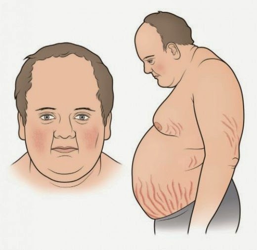
There are numerous signs and symptoms of Cushings along with a classic appearance:


Diagnosis
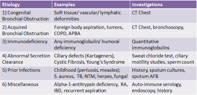
Guidelines recommend at least one first line test be performed to diagnose Cushing Syndrome - all of which assess cortisol levels (Endocrine society, 2008). If one of the tests is equivocal or the pre-test probability is high, another one of the first line tests should be performed. First line tests include one of the following:

1. Late night salivary cortisol - Elevated cortisol measurements suggestive of Cushing Syndrome.
2. 24 hr Urinary Free Cortisol - Elevated cortisol measurements suggestive of Cushing Syndrome.
3. Low-dose dexamethason suppression test - Assesses pituitary corticotroph response to steroids. Normal tissue 


Once a diagnosis of Cushing Syndrome is established, determing the underlying cause is the next step (ectopic ACTH production vs Pituitary ACTH production vs primary adrenal cortisol excess). Measuring a serum ACTH level is helpful, as it should be suppressed in primary adrenal tumors, but elevated in both ectopic and pituitary sources of Cushing Syndrome. 

If serum ACTH is elevated, the next step is to perform a High Dose Dexamethasone Suppression test. Pituitary sources of ACTH excess are only relatively resistant to negative feedack inhibition from glucocorticoid - therefore, with high doses of the potent dexamethasone, suppression of ACTH and therefore cortisol production is expected. However, ectopic sources of ACTH production will not be sensitive to negative feedback, and therefore persistently high cortisol levels are expected. See below.


One last thing...

Who's picture is that at the beginning of the post? That's Peter Cushing, who plays Grand Moff Tarkin in Star Wars: A New Hope (he's the commander of the Death Star). Any relation to Harvey Cushing, the first person to describe the disease? Nope. No relation.

References
Click here for the Endocrine 2008 guidelines on diagnosing Cushing

Sunday, October 5, 2014

Bronchiectasis

This past week, we discussed bronchiectasis in morning report with Dr. Shane Shapera. We had an interesting case of a young man with a history of recurrent sinus infections found to have hypogammaglobulinemia. Let's discuss this disease in detail.

Definition 
The suffix '-ectasis' refers to dilation of a tubular structure - in this case, the bronchii. For bronchiectasis to occur, two factors need to be present:
1. Infection
2. Impaired ability to clear the infection

These two features will help us generate a list of possible causes for this disease. Bronchiectasis is a chronic obstructive disorder that shares some similarities with COPD - obstructive pattern on PFT, recurrent inflammatory flares requiring hospitalization, and some benefit from inhaled bronchodilators.

Etiology
There is broad list of conditions that can lead to bronchiectasis:


Diagnosis
The diagnosis of bronchiectasis can be established on the basis of clinical symptoms and radiography alone. Classic symptoms include chronic cough and copious thick sputum production for months to years. Additional symptoms are less common, including shortness of breath, hemoptysis, pleuritic chest pain, and wheezing. These chronic symptoms are punctuated by recurrent episodes of infection requiring antibiotic therapy.

Radiologically, the classic findings on chest x-ray include linear atelectasis, dilated  airways which may appear as tram lines, and peripheral opacities representing mucopurulent plugs. On CT Chest, findings include airway dilation (1.5x greater in diameter than an adjacent vessel), bronchial wall thickening, tree-in-bud pattern due to mucopurulent plugs accompanied by post-obstructive air trapping in the small airways, and bronchial wall cysts.


Investigations
Aside from the above mentioned chest x-ray and CT chest, other important investigations include sputum cultures and pulmonary function testing. Outside of these, the remainder of the investigations should be targeted at determining the underlying etiology of bronchiectasis. See above for details.

Treatment
 Of all the causes of bronchiectasis, only a few have treatments that have shown benefit (some immunodeficiencies, nontuberculous mycobacterial infection, APBA, recurrent aspiration, rhematologic disease). Instead of aiming to treat the underlying cause, the treatment for bronchiectasis is targted at three things:
1. Controlling infection - Using antibiotics during acute flares that are tailored to prior sputum cultures is an important aspect of therapy. This may include coverage for Pseudomonas. Vaccination is also important in preventing future devastating infections.
2. Reducing inflammation - Using inhaled and systemic steroids in select patients during and in between exacerbations may help reduce secretions and wheezing. This may also include prolonged use of antibiotics for their anti-inflammatory effects (eg. macrolides) in those who have recurrent exacerbations.
3. Improving bronchial hygiene - For all patients, this is an important aspect of therapy that aims to control secretions. It includes pulmonary physiotherapy, postural drainage, 
Surgery of affected lung and transplantation is reserved for those who are refractory to medical treatment.

References:
Click here for the NEJM review from 2002.