Tuesday, December 2, 2014

Hypertension


Since we often discuss inpatient issues during morning report and noon rounds, let's focus on an outpatient issue instead. Hypertension (HTN) is extremely common (20% of Canadians over the age of 18) and is a signifcant risk factor for end-organ dysfunction. The inpatient management of hypertension is very different from the outpatient management of hypertension. The information below is from the CHEP Guidelines, a Canadian hypertension guideline.

Diagnosis
High blood pressure is defined by a BP >140/90. The flow chart for the diagnosis of HTN can be complication, but basically the diagnosis is often made over the course of 3 ambulatory clinic visits. HTN is defined by a BP greater than 140/90.

https://blogger.googleusercontent.com/img/b/R29vZ2xl/AVvXsEhlwu22UznjOsBx-gbZMd6ajD3En62W1u_Fl54mftV_UWDqiyU_TIwfqyGEQpaKzJWwkJKh2x0kM0a0F2tfQuNYettiW2SzrD5sJRS30gCQZnXhITYxcsZmjhYSXg1SSygWpIAiHJhl9J8/s1600/diagnosis+of+htn.PNG


The next step is to use either clinic BP monitoring (BPM), ambulatory monitoring (ABPM), or home monitoring. If using clinic BPM, a BP greater than 140/90 in three subsequent visits clinches the diagnosis, or a BP greater than 160/100 in any of those three diagnosis. For ABPM and Home BPM, a consistent BP of  greater than 135/85  makes the diagnosis.

Beware of two entities:
1. White Coat HTN: BP greater than 140 in the clinic but less than 135 via ABPM (should not treat as HTN).
2. Masked HTN: BP greater than 135 on APM but less than 140 in the clinic (should treat as HTN).

Investigations
Should be done for all patients with newly diagnosed HTN.
1. Urinalysis
2 .Blood chemistry (potassium, sodium and creatinine)
3. Fasting glucose and/or glycated hemoglobin (A1c)
4. Fasting total cholesterol and high density lipoprotein cholesterol (HDL), low density lipoprotein cholesterol (LDL), triglycerides.
5. Standard 12-leads ECG
                                                   
Treatment
1) Non-Pharmacological
o    Physical Exercise – Should be recommended 30-60 minutes of moderate intensity exercise (walking, jogging, cycling, or swimming) x4-7/week
o    Weight loss - BMI should be 18.5 to 24.9 kg/m²
o    Alcohol Consumption -  less than 2 drinks per dayno more than 14 drinks/week for men and 9/week for women
o    Salt Intake – Restrict to 2,000 mg (5g of salt or 87mmol of sodium) per day
o    Stress - Keep it down! Don't make me come over there.

2) Pharmacological
The first step is to remove agents that can induce/aggravate HTN (NSAIDs, steroids, OCP, EPO, MAOIs, SSRIs). Next, decide on a target for BP, which are listed below:

https://blogger.googleusercontent.com/img/b/R29vZ2xl/AVvXsEjM4ggFXRDq5cgHLkU5YLamWY-e_7zcMyucop66R8TpQJa7Y1Zb_bVx4BOlHfrVUBHeLDGFfn3Q4LOfCHySfi0SjIw-6BgsUFbtU4lTLSJaNbJqcheEed_uAImMGGr-zXrG6HnVrFD3unA/s1600/BP+Targets.PNG

Next, select an anti-HTN agent. Important considerations for choosing HTN treatment:
  • Use caution when initiating therapy with 2 drugs in whom adverse events are more likely (e.g. frail elderly, those with postural hypotension or who are dehydrated).
  • If a diuretic is not used as first or second line therapy, triple therapy should include a diuretic, when not contraindicated. 
  • ACE inhibitors, renin inhibitors, and ARBs are contraindicated in pregnancy
  • Don’t use an ACE inhibitor and an ARB together, except only in people with advanced heart failure or proteinuric nephropathy. This combination does not reduce cardiovascular events more than the ACEI alone and have more adverse effects.
  • Two drug combinations of beta blockers, ACE inhibitors, and angiotensin receptor blockers have not been proven to have additive hypotensive effects. Therefore, use if together only if non-blood pressure lowering indication.
  • ACE-inhibitors are not recommended (as monotherapy) for black patients without another compelling indication.

Here is the treatment algorithm for systolic/diasltolic HTN. Note that the algorithm is similar for Isolated Systolic HTN, except beta blockers and ACE inhibitors are not considered first line.

https://blogger.googleusercontent.com/img/b/R29vZ2xl/AVvXsEiroPDGLQUutWXLx1z_EbOxS-_MtNlHrgipgq1B69tQJNXArQegRfXF3D-QVICvRqaEGCKXB7giflLIuWNNvPRiTN4K4yg7lHKMli7UfTeHjEk4W0CKUjn4-O8g_N3kPszWHndUUVxbMtY/s1600/First+line+agents.PNG

Phewf. That was a lot of stuff.
Why a picture of Wonder Woman at the top? Turns out, the same person who created Wonder Woman also invented the polygraph test. Hence the 'lasso of truth.' Doesn't have much to do with hypertension, except that liars have higher blood pressure....probably.

An excellent resource is  https://www.hypertension.ca/en/chep. There, you can find all the guidelines for HTN diagnosis and treatment.


Tuesday, November 18, 2014

Metabolic Acidosis and Alkalosis


A common problem faced by trainees in Internal Medicine is acid-base disorders. Here, we will discuss the causes of acid-base disorders rather than the diagnosis on the basis of arterial blood gases. Specifically, we will discuss metabolic disturbances.

1) How does potassium affect acid-base status?
Changes in potassium often affect the metabolic acid/base status and vice versa. This is explained by the movement of excess potassium into cells which is balanced in part by intracellular hydrogen ions moving out to the extracellular fluid, and vice versa. This is termed "Internal Potassium Balance:" 


Therefore:
Metabolic acidosis tends to be associated with hyperkalemia (exceptions below).
Metabolic alkalosis tends to be associated with hypokalemia (exceptions below).

2) How does volume status affect acid-base status?
Hypovolemia can predispose patients to contraction alkalosis in some cases. There are a few reasons for this:
1. Loss of large volumes of low-bicarbonate, sodium-containing fluid leads to bicarbonate concentration rises because of contraction of the ECF volume with a constant quantity of  bicarbonate. This often occurs with IV loop diuretics.
2. High aldosterone - In the cortical collecting duct, high aldosterone released due to volume contractions leads to potassium and hydrogen ion excretion to balance sodium reabsorption. Hydrogen ion loss causes alkalosis, as does hypokalemia itself for the reasons stated above.
3. Angiontensin II - Released as part of RAAS activation in hypovolemia, angiontensin II acts at the Na+/H+ exchanger in the proximal tubules to enhance sodium absorption and H+loss, leading to alkalosis.

Therefore, contraction alkalosis can be seen in some patients with volume depletion.

Metabolic Acidosis

A) Increased Anion Gap
In these cases, extra anions (paired with H+) are introduced to the blood. The His consumed by HCO3- in the buffer system and the only component that remains is the anion, which creates the anion gap. One mnemonic that can be used to remember these causes is MUDPILES:


Methanol
Uremia (chronic kidney disease)
Diabetic Ketoacidosis
Propylene glycol/Paraldehyde
Iron overload/Isoniazid use
Lactic acidosis
Ethylene glycol
Salicylates 

B) Non-Anion Gap
In these cases, no new anions are being introduced to the blood. Instead, either HCO3-  is excessively lost or renal acid is not being excreted. 

Note that "hyperchloremic metabolic acidosis" is interchangeable with "non-anion gap metabolic acidosis." This is because chloride is proportionately increased when HCO3-  is lost or H+ accumulates (no extra anions are present).

Causes:
1. Diarrhea - Loss of bicarbonate rich fluid results in acidosis. It is  important to note that although diarrhea typically causes a metabolic acidosis, it can also cause a metabolic alkalosis in the following cases:
- Laxative abuse
- Villous adenoma
- Congenital chloroidorrhea (rare) 

2. Bowel conduits for urine - These conduits are created during ureteric or bladder operations. When urine is exposed to gut mucosa, it reabsorbs chloride in exchange for HCO3-, leading to acidosis.

3. Renal Tubular Acidosis
These diseases result from the kidney's inability to acidify the urine due to several reasons. Note that potassium and acid/base disturbance associations in these cases do not follow the usual pattern listed above.

Metabolic Alkalosis

Metabolic alkalosis results from additional creation of bicarbonate combined with a process which prevents renal excretion of the excess bicarbonate. Patients with metabolic alkalosis compensate by hypoventilating and should raise the PCO2 by about 0.7 mm Hg for every 1 meq/L elevation in the bicarbonate. This initially lowers the arterial pH toward normal and is most effective acutely, but then becomes less effective over time.

The differential diagnosis for metabolic alkalosis can be broken down into two groups, according to the urinary chloride levels:


Of these, the two most common causes are vomiting and diuretic therapy. One additional differential diagnosis for metabolic alkalosis of relevance is contraction alkalosis, mentioned earlier.

Grrrrrrrrrrrrrreat.
Here's a more physiological approach to acid-base disturbances from NEJM.

Thursday, November 6, 2014

Malaria

During morning report this week, we discussed a patient who returned form Ghana with Falciparum malaria. Let's discuss the approach to treatment of malaria - which literally means 'bad air.'


Malaria - What's up with that? 
Malaria is caused by the protozoan PlasmodiumThe mosquito that carries Plasmodium species called Anopheles (literally translated to 'ueseless'). Due to their transmission of malaria, mosquitos are thought to have caused half of all human deaths - more than all the wars combined.

Malaria should be suspected in anyone with a fever that has had mosquito exposure in a region where malaria is endemic. Don't expect to get a history of 'mosquito bites' from patients, just assume that they got mosquito bites when they traveled in the area. The incubation period for malaria: can range from 2 weeks (often falciparum) to a year, due to potential latency of the organisms. The diagnosis of malaria is based on light microscopy of thick and thin smears of patient blood - which are highly sensitive for detection of parasites when the load is >1%. When the parasite load is low, antigen testing is able to detect infection, though antigen testing cannot reveal the quantity of organisms infecting a patient. Also, it may or not be able to differentiate falciparum species from non-falciparum species.


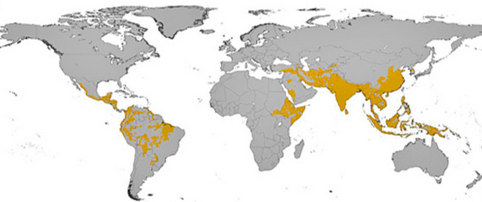
The top map shows falciparum distribution and the bottom shows vivax distribution.

4 Malaria Questions to consider for treatment in a patient diagnosed with Malaria:
1) Is this uncomplicated or complicated malaria?
2) Where it was acquired, and what are the resistance patterns there?
3) What species of malaria is it?
4) What is the degree of parasitemia?

1) Presentation of Malaria: Signs vs Symptoms

Uncomplicated vs Complicated Presentation of Malaria

In general, patients who are not obtunded and can take PO medications are considered "uncomplicated".

Uncomplicated Malaria - Usually presents with non-specific symptoms, such as chills, fatigue, diaphoresis, headache, nausea/vomiting, abdominal pain, diarrhea, arthralgias, and myalgias

On physical examination, the most prominent and common findings are splenomegaly and jaundice. This may be found even in patients without complicated malaria. Common lab abnormalities in patients with uncomplicated malaria include thrombocytopenia (very common) and anemia. Elevated liver enzymes, elevated creatinine, and raised WBC are also seen.
Complicated Malaria

  • Altered consciousness +/- seizures
  • Respiratory distress or acute respiratory distress syndrome (ARDS)
  • Circulatory collapse (shock)
  • Metabolic acidosis 
  • Renal failure, hemoglobinuria ("blackwater fever")
  • Liver failure
  • Coagulopathy with or without disseminated intravascular coagulation
  • Severe anemia or massive intravascular hemolysis
  • Hypoglycemia

2) Location of Acquisition 
Location of acquisition is relevant to understanding whether the organism is expected to be resistant or not. When in doubt, treat all falciparum organisms as if they are resistant to chloroquine. Chloroquine sensitive areas include Central America, Haiti, the Dominican Republic, former Soviet Union nations, and most of the Middle East. Anywhere outside of these regions is considered to be chloroquine resistant. Potential areas of resistance to artesunate include southeast Asia (especially Cambodia).


3) Species of Malaria


  • Non-falciparum (P. vivax, P. ovale, P. malariae, P. knowlesi) cause febrile illnesses but are rarely fatal.
  • However, P. falciparum infections are more virulent and can rapidly progress to fatal illness.
  • It is often impossible to determine the species of malaria infection on clinical grounds alone. This can be established through antigen testing and can sometimes be established based on the location of acquisition

    4) Level of Parasitemia 
    This refers to the percentage of RBCs carrying the organism and is established by examining the blood smear. In uncomplicated malaria, loads tend to be >5% in low transmission areas and 10% in high transmission areas.


    Treatment
    With chloroquine sensitive strains (determined by location of acquisition), the first line of therapy is either hydroxycloroquine or chloroquine. Side effects of these medications include GI upset, retinopathy, and pruritus.

    With chloroquine resistant strains, first line therapy usually includes Artesunate based therapy or Atovaquone-based therapy (+proguanil). Artesunate therapy is generally very well tolerated.

    What's that picture of the top? That's the building of the Panama Canal, 1904-1914. Malaria and yellow fever were rampant during the construction. Overall, 5600 people died building the canal.


    An excellent resource for malaria, as indicated by Dr. Paul Bunce:


    Wednesday, October 22, 2014

    Cushing Syndrome


    Cushing Syndrome (note that it should not be referred to as Cushing's Syndrome - the possessive form for eponyms is discouraged. Also, the use of eponyms is discouraged) describes the signs and symptoms associated with prolonged cortisol excess of any cause. Cushing Disease specifically refers to cortisol excess due to pituitary stimulation by ACTH. Let's discuss the diagnosis of Cushing Syndrome. First, the presentation of Cushing.

    Signs and Symptoms
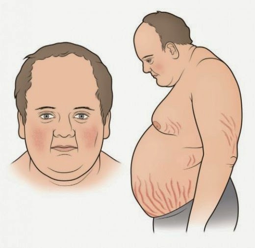
    There are numerous signs and symptoms of Cushings along with a classic appearance:


    Diagnosis
    Guidelines recommend at least one first line test be performed to diagnose Cushing Syndrome - all of which assess cortisol levels (Endocrine society, 2008). If one of the tests is equivocal or the pre-test probability is high, another one of the first line tests should be performed. First line tests include one of the following:

    1. Late night salivary cortisol - Elevated cortisol measurements suggestive of Cushing Syndrome.
    2. 24 hr Urinary Free Cortisol - Elevated cortisol measurements suggestive of Cushing Syndrome.
    3. Low-dose dexamethason suppression test - Assesses pituitary corticotroph response to steroids. Normal tissue 


    Once a diagnosis of Cushing Syndrome is established, determing the underlying cause is the next step (ectopic ACTH production vs Pituitary ACTH production vs primary adrenal cortisol excess). Measuring a serum ACTH level is helpful, as it should be suppressed in primary adrenal tumors, but elevated in both ectopic and pituitary sources of Cushing Syndrome. 

    If serum ACTH is elevated, the next step is to perform a High Dose Dexamethasone Suppression test. Pituitary sources of ACTH excess are only relatively resistant to negative feedack inhibition from glucocorticoid - therefore, with high doses of the potent dexamethasone, suppression of ACTH and therefore cortisol production is expected. However, ectopic sources of ACTH production will not be sensitive to negative feedback, and therefore persistently high cortisol levels are expected. See below.


    One last thing...

    Who's picture is that at the beginning of the post? That's Peter Cushing, who plays Grand Moff Tarkin in Star Wars: A New Hope (he's the commander of the Death Star). Any relation to Harvey Cushing, the first person to describe the disease? Nope. No relation.

    References
    Click here for the Endocrine 2008 guidelines on diagnosing Cushing

    Sunday, October 5, 2014

    Bronchiectasis

    This past week, we discussed bronchiectasis in morning report with Dr. Shane Shapera. We had an interesting case of a young man with a history of recurrent sinus infections found to have hypogammaglobulinemia. Let's discuss this disease in detail.

    Definition 
    The suffix '-ectasis' refers to dilation of a tubular structure - in this case, the bronchii. For bronchiectasis to occur, two factors need to be present:
    1. Infection
    2. Impaired ability to clear the infection

    These two features will help us generate a list of possible causes for this disease. Bronchiectasis is a chronic obstructive disorder that shares some similarities with COPD - obstructive pattern on PFT, recurrent inflammatory flares requiring hospitalization, and some benefit from inhaled bronchodilators.

    Etiology
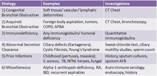
    There is broad list of conditions that can lead to bronchiectasis:


    Diagnosis
    The diagnosis of bronchiectasis can be established on the basis of clinical symptoms and radiography alone. Classic symptoms include chronic cough and copious thick sputum production for months to years. Additional symptoms are less common, including shortness of breath, hemoptysis, pleuritic chest pain, and wheezing. These chronic symptoms are punctuated by recurrent episodes of infection requiring antibiotic therapy.

    Radiologically, the classic findings on chest x-ray include linear atelectasis, dilated  airways which may appear as tram lines, and peripheral opacities representing mucopurulent plugs. On CT Chest, findings include airway dilation (1.5x greater in diameter than an adjacent vessel), bronchial wall thickening, tree-in-bud pattern due to mucopurulent plugs accompanied by post-obstructive air trapping in the small airways, and bronchial wall cysts.


    Investigations
    Aside from the above mentioned chest x-ray and CT chest, other important investigations include sputum cultures and pulmonary function testing. Outside of these, the remainder of the investigations should be targeted at determining the underlying etiology of bronchiectasis. See above for details.

    Treatment
     Of all the causes of bronchiectasis, only a few have treatments that have shown benefit (some immunodeficiencies, nontuberculous mycobacterial infection, APBA, recurrent aspiration, rhematologic disease). Instead of aiming to treat the underlying cause, the treatment for bronchiectasis is targted at three things:
    1. Controlling infection - Using antibiotics during acute flares that are tailored to prior sputum cultures is an important aspect of therapy. This may include coverage for Pseudomonas. Vaccination is also important in preventing future devastating infections.
    2. Reducing inflammation - Using inhaled and systemic steroids in select patients during and in between exacerbations may help reduce secretions and wheezing. This may also include prolonged use of antibiotics for their anti-inflammatory effects (eg. macrolides) in those who have recurrent exacerbations.
    3. Improving bronchial hygiene - For all patients, this is an important aspect of therapy that aims to control secretions. It includes pulmonary physiotherapy, postural drainage, 
    Surgery of affected lung and transplantation is reserved for those who are refractory to medical treatment.

    References:
    Click here for the NEJM review from 2002.