Monday, July 9, 2012
CMA Driving Guidelines
In addition to the discussion below about jolt accentuation and other tests for meningeal irritation, check out the CMA's driving guidelines.
We discussed patients who have had a TIA- as you can read on this website there is no clear time period during which patients cannot drive- they need to be seen by a physician and if no neurological deficits persist can go back to driving.
Patients with a stroke should not drive for at least one month- however how long exactly depends on the physician's assessment of their deficits and risk of recurrence.
This website is a great resource as it is nearly impossible to remember all the times for every condition you will encounter. http://www.cma.ca/determining-fitness-to-drive

How are your meninges?
Today in morning report we discussed a case of a patient with fever, headache, and focal neurological signs. Those in the room were appropriately concerned about a CNS cause.
Could this patient have meningitis or something else (such as blood in subarachnoid hemorrhage) irritating his meninges?
Symptoms suggestive of meningeal irritation are: headache, neck stiffness, photophobia, cranial nerve palsies. These, in addition to fever/chills and other infectious symptoms can suggest a diagnosis of meningitis.
In meningitis, clinical history does a poor job of leading us to the diagnosis. Neck stiffness on history has a sensitivity of only 28%.
For physical exam, Kernig's and Brudzinski's signs have poor sensitivity and specificity (based on little data) for meningitis.
Jolt accentuation of headache is quite sensitive (97%) based on one study of 54 patients with fever and headache, making it a good ruling-out test if it is negative. To perform the test, ask the patient to shake their head from side-to-side at about 2-3Hz and ask them if their headache is better, the same or worse. If it is not worse, this significantly decreases the likelihood of meningitis.
It generates a positive likelihood ratio of 2.4 and negative likelihood ratio of 0.05 for CSF pleiocytosis (increased cell count). In order to use a likelihood ratio, you multiply the pre-test probability by the LR to get the post-test probability. See the image below for a visual of this calculation:
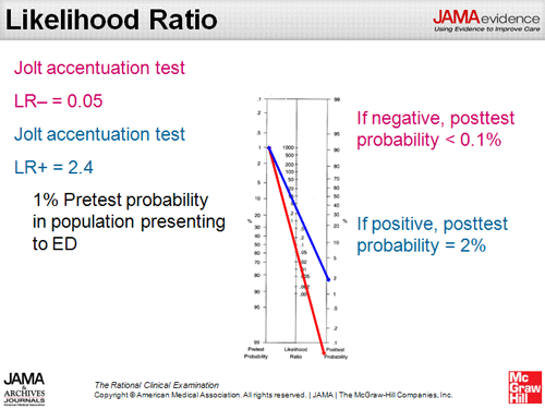
You can read more about this in the JAMA Rational Clinical Exam Article "Does This Patient Have Acute Meningitis" follow link here http://jama.jamanetwork.com/article.aspx?articleid=774331#JRC80004T4
Friday, July 6, 2012
Renal Disease in Multiple Myeloma
Today we talked about a woman with acute kidney injury in the setting of multiple myeloma. Renal impairment is seen in 20-40% of newly diagnosed patients with myeloma.
There are many ways that MM can damage a kidney:
1) Pre-renal:
- Hypercalcemia via renal vasoconstriction and volume depletion
2) Renal Glomerular:
- Amyloid deposits in glomerulus
- Light Chain Deposition Disease (LCDD)
- rarely Type 1 cryoglobulinemia
3) Renal Tubulo-Interstitial
- Tubular Cast (aggregates of light chains and Tamm-Horsfall protein) formation- "Myeloma Kidney"
- Light chain toxicity to renal tubules
- Acquired Fanconi syndrome (also a form of light chain toxicity to the proximal tubule)- usually asymptomatic but detected because of hypouricemia
- Interstitial plasma cell infiltration
- Chronic urate nephropathy
One key question is whether or not a multiple myeloma patient with renal failure requires a kidney biopsy. If light chains are high in the urine, myeloma kidney is the likely cause. In a setting where 24-hour urine collection shows nephrotic syndrome but little to no light chains, a biopsy is useful to make the definitive diagnosis of amyloid or light-chain related glomerular disease.
Tubular cast on path:

Thursday, July 5, 2012
Pemphi-What?
Today in morning report we heard about a patient with probable bullous pemphigoid. The pemphigoid/pemphigus question is a favourite on exams, and it can be a little confusing remembering the differences.
Pemphigus vulgaris is more serious because:
- loss of cohesion between cells leads to acantholysis, with flaccid, easily shearable bullae (and + Nikolsky sign)-> this leads to more denuded skin and thus more infections
- involves mucous membranes
Bullous pemphigoid= tense bullae (don't always break)- see pic below
Here
is a system to remember the characteristics of these two autoimmune skin
conditions:
Pemphigus =
Serious (thank you Grace for this trick!)
Pemphigus vulgaris is more serious because:
- loss of cohesion between cells leads to acantholysis, with flaccid, easily shearable bullae (and + Nikolsky sign)-> this leads to more denuded skin and thus more infections
- involves mucous membranes
- antibodies are to basement membrane, cells can still stick together
- usually seen in the elderly

- mucosal involvement less common
Definitive diagnosis of the two conditions relies on direct immunoflorescence microscopy of a biopsy sample to see where antibodies are located (epidermal basement membrane versus cell surface).
See you tomorrow!
- usually seen in the elderly

- mucosal involvement less common
Definitive diagnosis of the two conditions relies on direct immunoflorescence microscopy of a biopsy sample to see where antibodies are located (epidermal basement membrane versus cell surface).
See you tomorrow!
Wednesday, July 4, 2012
Tuesday, July 3, 2012
Welcome
Hello and welcome. I just started as the new chief resident, and am looking forward to posting some good stuff for you! Stay tuned.
Friday, April 27, 2012
Pleuritic Chest Pain - Friday April 27, 2012
Thank you to Dr.
Ho-Ping Kong for hosting today’s morning report, and thank you to team 7 for
bringing the case.
Today we
discussed a case with a man (in his 40’s) who presented with pleuritic chest
pain. We discussed the differential
diagnosis of pleuritic chest pain that gets worse when lying down and improves
with sitting up: pulmonary embolism,
pneumonia, pericarditis. Also, given the
nature of his pain, pancreatitis and GERD can sometimes have these symptoms,
too. This patient also had a history of
upper respiratory tract infection few weeks prior. ECG showed changes compatible with
pericarditis (with negative troponin).
These changes may include diffuse ST elevation that is concave up
(except for V1 and aVR) and PR segment depression.
We attributed
his pericarditis to a viral illness. One
of the common viral causes is coxsackie virus (although we really cannot prove
this). We also discussed other causes of
pericarditis include: infection (viral,
bacterial, TB, rarely fungal), transmural MI, after CABG, connective tissue
disease (e.g. lupus), uremia, drugs, chest wall radiation, chest wall trauma, and
neoplastic disease.
As always, we
also learned some medical trivia along the way.
Banting and Best treated their first patient with insulin at Toronto
General Hospital, it was a patient from the United States. When someone has pneumonia and GI symptoms
(e.g. diarrhea), it is important to consider Legionella as a causative
organism.
You can find out
more about ECG changes of pericarditis at ECG Made Simple (linked here). It is a very good resource for learning many
things about ECG. You can also read more
about pericarditis in this article (linked here).
Thank you everyone
for a great month! I have thoroughly
enjoyed working with all of you. Chris
Smith will be back Monday.
Tuesday, April 24, 2012
Anaphylaxis - Tuesday April 24, 2012
Thank you to Dr.
A. Page for hosting morning report and to team 5 for bringing a case from
overnight.
We discussed the
diagnosis and management of someone presenting with anaphylaxis. The diagnosis is made by meeting one of the
following minutes to hours after exposure to a likely allergen:
- skin/mucosal
manifestation + 1 of shock (decreased BP), or respiratory compromise.
- Two or more
of: skin/mucosal manifestation,
respiratory symptoms, hypotension, or GI symptoms.
- Hypotension after
exposure of an exposure to a known allergen for that patient.
We discussed the
management of anaphylaxis which include
- Securing
airway
- Obtaining IV
access
- Epinephrine
injection (0.3 mg IM)
- IV fluid
- Benadryl (diphenhydramine)
50 mg (H1 blocker)
- Ranitidine 50
mg (H2 blocker)
- Methyprednisolone
125 mg IV
- Bronchodilators
may be used
We also
discussed that there can be a biphasic reaction hours after the initial
episode.
We also
discussed that for patients on beta-blocker, epinephrine may be ineffective,
and that glucagon is needed.
Friday, April 20, 2012
Grand Morning Report - Friday April 20, 2012
Thank you to Dr.
Y. Patel for hosting Grand Morning Report.
Last night, we had 10 cases, but they had a few themes in common.
The first theme
is GI bleeding. We discussed that stools
can be black for a number of reasons (including iron supplementation). It is important to decide whether the patient
is having melena (by a digital rectal exam) and also to elucidate the temporal
relation of dark stool to other events (such as beginning of iron
supplementation). We also discussed that
fecal occult blood test is meant for a screening test for colon cancer and that
its utility in a bleeding patient is not tested. We also discussed the importance of
establishing good IV access.
We also
discussed when to transfuse. There is a
recently published clinical practice guideline in the Annals of Internal
Medicine (March 26, 2012) (linked here) on transfusion threshold for
hemoglobin. For patients who are not
bleeding and stable (without ACS), transfusion threshold of 70-80 is commonly
used. However, for bleeding patient, it
depends on patient stability, symptoms, whether bleeding is on-going, etc …
We then moved on
to the second theme of stroke. We
discussed that there are investigations that are sometimes helpful in patients
presenting with a stroke, but not necessarily in ALL patients. We must understand the purpose of each test
before ordering. For example, an MRI
brain is sometimes ordered to confirm the diagnosis of strokes (for example, in
the posterior circulation). However, if
the diagnosis is already very clear (e.g. on CT imaging), this may not be
necessary. Also, echocardiogram is used
to look for source for cardio-embolic stroke.
If a patient is known to have atrial fibrillation and is already
anti-coagulated, and that anticoagulation is planned to continue when safe to
do so, there may not be a need for an echocardiogram as it does not change
management. We also discussed that
carotid endarterectomy for symptomatic carotid artery stenosis has
evidence-based support. However, the
benefit for asymptomatic disease is marginal.
See this review in the Mayo Clinic Proceedings (linked here).
Thursday, April 19, 2012
Shortness of breath on exertion and pulmonary embolism - Thursday April 19, 2012
Thank you to
team 5 for bringing an interesting case.
Today, we
discussed a case of a man with significant previous coronary artery disease
treated with coronary artery bypass graft surgery and multiple stenting. He has known
LVEF of 20-39%. He presented to the
Emergency Department with shortness of breath on exertion.
We discussed the
pertinent history features when someone presents with previous coronary artery
disease. They include previous
infarctions, interventions (surgery, interventional, or medical), presence of
heart failure, and functional status (CCS class). We also discussed the reason for dual
anti-platelet therapy post-stenting and the minimum duration required for this
therapy. We discussed some of the
typical medical management for patients with coronary artery disease
(anti-platelet, beta-blocker, ACE-inhibitor, statin). We compared this with the medications that
our patient was on. We then generated a
list of differential diagnoses for our patient, which included heart failure
(exacerbation), pulmonary embolism, pneumonia, new ischemia, bleeding, and less
likely obstructive lung disease, or malignancy.
We also discussed the etiologies of heart failure and its triggers.
We looked at his
CXR, which showed poor inspiration (we discussed how to determine this by
counting ribs), with a right-sided consolidation. The CT thorax showed pulmonary embolism
(likely not acute) and right-sided consolidation. Five months ago, this patient had a “high
probability” V/Q scan followed by a negative CT PE study at a different
hospital. The team started this patient
on anticoagulation.
We then
discussed the etiology of his PE. We
discussed the usual risk factors for PE such as trauma, surgery, malignancy,
immobility (including travel), and thrombophilia. We did not have enough time to discuss this
thoroughly, but we did note that this patient developed new-onset microcytic
anemia and that a work-up is necessary.
Wednesday, April 18, 2012
Shortness of breath in a young person - Wednesday April 18, 2012
Thank you to Dr.
P. Bunce for hosting today’s morning report and to team 7 for bringing a case.
We discussed a
case of a previously healthy young person who presented with shortness of
breath. We discussed a list of
differential diagnoses of this problem in a young person that is different than
the one we usually use for an elderly person presenting with the same complaint. The reason we did this is given that she is
previously healthy, diagnoses such as heart failure is less likely in the
absence of hypertension, diabetes or coronary artery disease. For our patient, the differential diagnoses
constructed were: asthma, pneumonia, pulmonary embolism, malignancy, pregnancy,
pulmonary hypertension. We also discussed
that TB is a consideration depending on risk factors and exposure history.
We then looked
at her CXR and CT of thorax and it showed an anterior mediastinal mass, and
pulmonary embolism. We discussed the
differential diagnoses of T’s. Teratoma,
thymoma, thyroid, and terrible lymphoma.
Occasionally, TB can also be a cause.
Tissue was obtained.
Tuesday, April 17, 2012
Hemochromatosis and Febrile Neutropenia - Tuesday April 17, 2012
Thank you Dr. A.
Page for hosting morning report and to team 8 for bringing a fascinating case.
Today, we
discussed a patient with hemochromatosis and many of its complications
including liver cirrhosis, heart failure, diabetes, hypothyroidism, and
hypogodnadism. This patient was also
started on oral iron chelation therapy (deferiprone) and presented with febrile
neutropenia with a 1-day history of fever, malaise, dry cough, enlarged
cervical lymph nodes, dysphagia, and rash on trunk.
We first
discussed hemochromatosis as an autosomal recessive genetic disease (most
common mutation is C282Y on the HFE gene) that results in iron overload. Patients have high iron saturation (>45%)
and high ferritin. Genetic testing is
done for diagnosis in the presence of iron overload. Iron deposits can cause dysfunction in many
organs including liver, heart, pancreas (diabetes), skin, gonads, and
joints. Main treatment is phlebotomy
and/or iron chelation (which our patient is on).
We discussed the
etiology of the patient’s neutropenia is likely secondary to deferiprone (known
to cause agranulocytosis). We also
discussed other causes of neutropenia including bone marrow suppression from
infection or drugs (e.g. levamisole, chemotherapy, …), nutritional deficiency,
and infiltration of bone marrow. He
would be treated like any other patients with febrile neutropenia requiring
empiric broad-spectrum antibiotics coverage until organism/source identified
and/or recovery of neutrophil counts.
We also
discussed the presentation of dry cough, enlarged lymph nodes, dysphagia, and
malaise. While a non-infectious cause
(e.g. lymphoma) is possible, the onset is quite rapid. We focused our discussion on infectious
causes that included: bacterial (group A
strep, Arcanobacerium haemolyticum), viruses (HIV, hepatitis, EBV, CMV, Measle,
Parvo virus), fungal disease (mucormycosis, less likely Candida), and TB. We also discussed some syndromes involving
infection of the head and neck area including Ludwig’s Angina, Lemierre’s syndrome
(septic thrombophlebitis, most common Fusobacterium), and Vincent angina.
Subscribe to:
Posts (Atom)