Wednesday, October 22, 2014

Cushing Syndrome


Cushing Syndrome (note that it should not be referred to as Cushing's Syndrome - the possessive form for eponyms is discouraged. Also, the use of eponyms is discouraged) describes the signs and symptoms associated with prolonged cortisol excess of any cause. Cushing Disease specifically refers to cortisol excess due to pituitary stimulation by ACTH. Let's discuss the diagnosis of Cushing Syndrome. First, the presentation of Cushing.

Signs and Symptoms
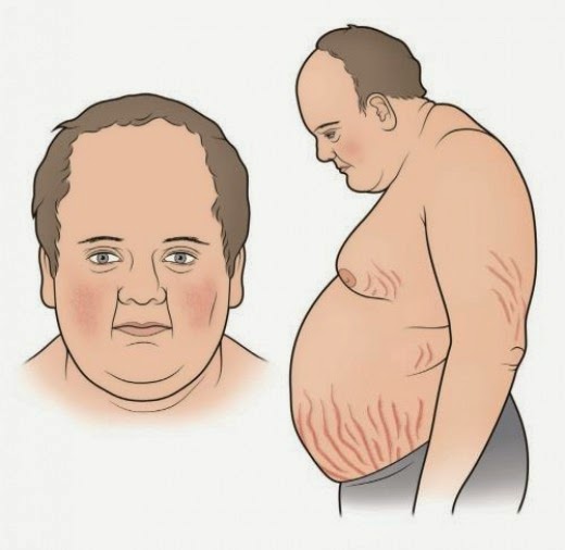
There are numerous signs and symptoms of Cushings along with a classic appearance:



Diagnosis
Guidelines recommend at least one first line test be performed to diagnose Cushing Syndrome - all of which assess cortisol levels (Endocrine society, 2008). If one of the tests is equivocal or the pre-test probability is high, another one of the first line tests should be performed. First line tests include one of the following:

1. Late night salivary cortisol - Elevated cortisol measurements suggestive of Cushing Syndrome.
2. 24 hr Urinary Free Cortisol - Elevated cortisol measurements suggestive of Cushing Syndrome.
3. Low-dose dexamethason suppression test - Assesses pituitary corticotroph response to steroids. Normal tissue 


Once a diagnosis of Cushing Syndrome is established, determing the underlying cause is the next step (ectopic ACTH production vs Pituitary ACTH production vs primary adrenal cortisol excess). Measuring a serum ACTH level is helpful, as it should be suppressed in primary adrenal tumors, but elevated in both ectopic and pituitary sources of Cushing Syndrome. 

If serum ACTH is elevated, the next step is to perform a High Dose Dexamethasone Suppression test. Pituitary sources of ACTH excess are only relatively resistant to negative feedack inhibition from glucocorticoid - therefore, with high doses of the potent dexamethasone, suppression of ACTH and therefore cortisol production is expected. However, ectopic sources of ACTH production will not be sensitive to negative feedback, and therefore persistently high cortisol levels are expected. See below.


One last thing...

Who's picture is that at the beginning of the post? That's Peter Cushing, who plays Grand Moff Tarkin in Star Wars: A New Hope (he's the commander of the Death Star). Any relation to Harvey Cushing, the first person to describe the disease? Nope. No relation.

References
Click here for the Endocrine 2008 guidelines on diagnosing Cushing

Sunday, October 5, 2014

Bronchiectasis

This past week, we discussed bronchiectasis in morning report with Dr. Shane Shapera. We had an interesting case of a young man with a history of recurrent sinus infections found to have hypogammaglobulinemia. Let's discuss this disease in detail.

Definition 
The suffix '-ectasis' refers to dilation of a tubular structure - in this case, the bronchii. For bronchiectasis to occur, two factors need to be present:
1. Infection
2. Impaired ability to clear the infection

These two features will help us generate a list of possible causes for this disease. Bronchiectasis is a chronic obstructive disorder that shares some similarities with COPD - obstructive pattern on PFT, recurrent inflammatory flares requiring hospitalization, and some benefit from inhaled bronchodilators.

Etiology
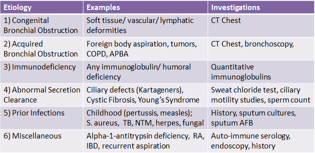
There is broad list of conditions that can lead to bronchiectasis:


Diagnosis
The diagnosis of bronchiectasis can be established on the basis of clinical symptoms and radiography alone. Classic symptoms include chronic cough and copious thick sputum production for months to years. Additional symptoms are less common, including shortness of breath, hemoptysis, pleuritic chest pain, and wheezing. These chronic symptoms are punctuated by recurrent episodes of infection requiring antibiotic therapy.

Radiologically, the classic findings on chest x-ray include linear atelectasis, dilated  airways which may appear as tram lines, and peripheral opacities representing mucopurulent plugs. On CT Chest, findings include airway dilation (1.5x greater in diameter than an adjacent vessel), bronchial wall thickening, tree-in-bud pattern due to mucopurulent plugs accompanied by post-obstructive air trapping in the small airways, and bronchial wall cysts.


Investigations
Aside from the above mentioned chest x-ray and CT chest, other important investigations include sputum cultures and pulmonary function testing. Outside of these, the remainder of the investigations should be targeted at determining the underlying etiology of bronchiectasis. See above for details.

Treatment
 Of all the causes of bronchiectasis, only a few have treatments that have shown benefit (some immunodeficiencies, nontuberculous mycobacterial infection, APBA, recurrent aspiration, rhematologic disease). Instead of aiming to treat the underlying cause, the treatment for bronchiectasis is targted at three things:
1. Controlling infection - Using antibiotics during acute flares that are tailored to prior sputum cultures is an important aspect of therapy. This may include coverage for Pseudomonas. Vaccination is also important in preventing future devastating infections.
2. Reducing inflammation - Using inhaled and systemic steroids in select patients during and in between exacerbations may help reduce secretions and wheezing. This may also include prolonged use of antibiotics for their anti-inflammatory effects (eg. macrolides) in those who have recurrent exacerbations.
3. Improving bronchial hygiene - For all patients, this is an important aspect of therapy that aims to control secretions. It includes pulmonary physiotherapy, postural drainage, 
Surgery of affected lung and transplantation is reserved for those who are refractory to medical treatment.

References:
Click here for the NEJM review from 2002.

Sunday, September 21, 2014

Graft Versus Host Disease: A TGH Special

During your time at TGH, you will likely encounter the entity of Graft Versus Host Disease (GVHD). It is  not a common disease; however, it is commonly seen at TGH due to our immune-suppressed patients, especially in transplanted patients. Let's first discuss the types of stem cell/bone marrow transplant (aka Hematopoietic cell transplantation).

Autologous: Hematopoietic cells are harvested from the bone marrow or peripheral blood prior to high-dose myeloablative chemotherapy/radiotherapy. The cells are then later reinfused back into the same patient during a period of prolonged bone marrow failure in order to help restore hematopoiesis. This type of transplant is used in lymphoma and myeloma, and has previously been used in patients with some solid tumors. 
Allogeneic: Stem cells are harvested from the bone marrow of healthy donors or newborn umbilical cord and are infused to reconstitute a host hematopoeitic system following myeloablative therapy. Allogenic stem cell transplants carry the same risks as autologous transplants, with the additional risk of graft-versus-host-disease.

Graft Versus Host Disease (GVHD)
GVHD occurs when donated lymphocytes attack normal host tissues because they are recognized as 'foreign.' GVHD is a common consequence of allogenic bone marrow transplantation, occurring in up to 40% of patients. It is a potential complication of all transplantation, including transfusion of blood products, but is particularly concerning in stem cell transplantations due to the inherent immune suppression of patient's undergoing stem cell or bone marrow transplantation. Due to their immune-suppressed nature, it is often difficult to differentiate infections from GVHD in patients who have received allogeneic transplants.

Risk Factors for GVHD
- Degree of HLA mismatching 
- Older age
- The use of peripheral blood/bone marrow samples instead of umbilical cord blood 

Sites Most Commonly Affected by GVHD
Skin - Lichen planus-like or morphea-like changes, sclerotic features. Diagnosis can be made without biopsy. 
Gut - Mucosal sloughing, severe cramps with secretory diarrhea. Diagnosis made on biopsy
Liver - Cholestatic hepatitis with nausea and vomiting.
Lungs - Bronchiolitis obliterans. Diagnosis made on biopsy.


Forms of GVHD
Acute
This form usually occurs before 100 days post-transplant and can present with a classic maculopapular or bullous rash, cholestasis with elevated bilirubin, nausea & vomiting, and abdominal cramps with diarrhea. Acute GVHD can occur more than 100 days after transplant, and is known as Late-Onset Acute GVHD.

Chronic
The chronic form more resembles other autoimmune disorders, presenting with arthritis, scleroderma-like skin changes or lichen planus, sicca syndrome, and chronic hepatitis with rising bilirubin. Patients may sometimes present with features of both chronic and acute GVHD, in which case they are labeled with an 'Overlap' syndrome.

Treatment
Because GVHD is immune-mediated, it is often treated with glucocorticosteroids. This therapy can be local (topical for skin, enteric for gut) or systemic (oral or IV). Other therapies include cyclosporine, MMF, and azathioprine. Complications related to the disease and its treatment must be monitored carefully.

Sunday, September 7, 2014

Hypertrophic Obstructive Cardiomyopathy

This week during morning report we discussed a young woman who presented with chest pain, positive serum troponin, and a known history of hypertrophic cardiomyopathy (HCM). Let's discuss the physical examination findings and management of this condition.

Physical Examination
Classic findings for HCM on physical examination are seen in patients with left ventricular outflow tract (LVOT) obstruction. Patients with none or small gradients may have no physical examination findings.

A. Auscultation:

LVOT Obstruction - This murmur is similar sounding to aortic stenosis, in that it is a systolic crescendo-decrescendo murmur heard across the precordium. However, a major distinction between the murmurs is that the HCM murmur increases with any maneuver that decreases pre-load, whereas aortic stenosis either decreases or is not affected by these maneuvers. This is because decreased pre-load leads to a smaller LV cavity and thus causes more dynamic obstruction during systole. See below for details on the difference between the murmur of HCM and aortic stenosis.


SAM (systolic anterior motion) - This murmur is heard best in the mitral area and is due to the in-drawing of the mitral leaflets during systole, causing a murmur that is similar in sound to mitral regurgitation. SAM contributes to LVOT obstruction and is an important physical exam finding.

S4 - An S4 is commonly heard in HCM, best auscultated using the bell of the stethoscope in the apex. This sound is heard in aortic stenosis only in advanced cases.

B. Other findings

- Paradoxical splitting of the S2 is heard when the LVOT obstruction is severe.
- The carotid upstroke is often brisk and bifid. Bifid means two upstrokes are palpable - the initial pulsation due to LV contraction, a softening of the pulse due to dynamic obstruction, and a second impulse at the end of systole as the ventricular pressure overcomes the obstruction.
- The apical pulse may also be bifid. When an S4 is also palpable, this is known as the rarely seen "triple ripple." In general, the apical pulse will be diffuse and strong.


Management

1. Non-medical - Patients with HCM should be counselled to avoid situations which cause peripheral vasodilation or dehydration. This includes avoiding strenuous exercise, alcohol intake, and diuretics if possible.

2. Pharmacological Therapy- In order to increase diastolic ventricular filling time (and thus increase pre-load), negative chronotropic medications are indicated in patients with HCM. This includes beta-blockers and nondihydropiridine calcium channel blockers. The next line of therapy is negative ionotropic therapy with disopyramide, which is also an anti-arrythmic medication.

3. Surgical Myomectomy - In patients who remain symptomatic despite medications, surgical therapy is indicated. The procedure involves excising the basal septum and is highly successful. It can cure SAM and therefore reduce LVOT obstruction, and is considered the gold standard operative management of HCM.

4. Alcohol Ablation - In this procedure, alcohol is injected into a septal perforating artery via coronary artery catheterization. It is a less invasive therapy than surgical myomectomy, but myomectomy has more proven long-term efficacy.

5. Implanted Defibrillator - Consideration for this therapy is based on assessment for risk of sudden cardiac death, including family history of sudden cardiac death, wall thickness, genetic defect, gradient of outflow obstruction, and history of arrhythmia. Patients with HCM are at-risk for arrythmias due to disorganized myocytes and relatively poorly perfused myocardial perforators due to thickened myocardium.


That's all, folks!

Here's the obligatory NEJM article on HCM.
Here's a review article by one of the pioneers of HCM research, Dr. Doug Wigle from TGH.


Sunday, August 24, 2014

Severe B12 Deficiency


During morning report this past week, we were introduced to a patient with severe alcoholism who presented with a hemoglobin of 24 g/L due to B12 deficiency, whose only presenting symptom was 'fatigue.' That's pretty low. Low enough to hear orbital bruits. It is important to note that it takes months to years to deplete one's Vitamin B12 stores as it did in this patient's case.
On physical examination, the patient appeared "pale and yellow," ( a lemony yellow) due to the combination of anemia and hemolysis, a classic appearance of patients with severe B12 Deficiency. Let's go over the important pieces of diagnosing and treating B12 deficiency:

Causes
To develop a differential diagnosis of B12 deficiency, think of the anatomic components necessary for B12 absorption, from mouth to terminal ileum:
Mouth (Diet) - Vegans, alcoholics, patients with eating disorders
Stomach - Pernicious anemia results from autoantibiodies to parietal cells due to Autoimmune Atrophic Gastritis. This is the most common cause of B12 deficiency in adults.
Proximal Small Bowel - Post Roux-en-Y, Celiac Disease, Chronic Pancreatitis,
Terminal Ileum - IBD is a disease with classic TI manifestations.

Physical Examination
Aside from the above mentioned finding of "pale and yellow," another important aspect of physical examination is looking for risk factors for B12 deficiency, such as manifestations of alcoholism, general malnutrition, IBD, and other autoimmune disease.

The neurological examination is unique in patients with B12 deficiency. It is one of the very few diseases that can manifest with both upper and lower neuron lesions. Manifestations classically include subacute combined degeneration of the dorsal (posterior) and lateral spinal columns. SCD manifests as symmetrical primarily lower limb neuropathy with loss of vibration and position sense, which can result in ataxia. Other neurologic findings in B12 deficiency include axonal degeneration of peripheral nerves and central nervous system symptoms including memory loss, irritability, and dementia. Notably, not all patients with neurologic abnormalities secondary to B12 deficiency have hematologic manifestations. 

Investigations
Aside from macrocytic anemia, the other findings on investigation that are suggestive of B12 deficiency include a low or undetectable serum B12 level, and megaloblastic anemia.

Blood films from patients with severe B12 deficiency will also show hemolysis, due to improper RBC production in the bone marrow. Low platelets may also be seen because B12 is necessary for the production of megakaryocytes. The finding of anemia, hemolysis, and thrombocytopenia on a blood film may be confused with a MAHA. The distinction is that the patients with B12 deficiency will also megaloblastic anemia:
- Megaloblasts (immature RBCs) in the bone marrow
- Hypersegmented neutrophils (>5  nuclear lobes) on their peripheral blood film,  


Important tests to help diagnose the etiology of B12 deficiency include antibodies to IF, the Schilling Test, and endoscopy.

Important Tips for treatment outlined during our discussion with Dr. Wayne Gold:

1) Use B12 Injections to Supplement B12 - Once a diagnosis of Vitamin B12 deficiency is made, use injection rather than oral supplements to replete patients. This will allow you to bypass the entire GI tract and will reliably provide patients with your supplement.

2) Watch out for Hypokalemia - When treating patients with severe B12 deficiency, new RBCs are being produced and numerous building blocks are consumed. This includes potassium, which is a major intracellular electrolyte. Remember to carefully replace patient's potassium during treatment.

3) Watch out for folate deficiency - As new RBCs are being produced, folate is rapidly consumed and so when patients are receiving B12 supplementation they will also require folate supplementation as well.

4) Don't transfuse too much - Patients with severe, chronic anemia compensate slowly over time with a hyperdynamic cardiac state, and will have no symptoms of hemodynamic instability. Over-treatment by physicians to rapidly transfuse these patients to a normal hemoglobin values can lead to cardiac failure due to volume overload in an already stressed cardiovascular system.

Here are some good review articles:
Click here for a good article on the treatment of B12 deficiency from Blood.
And a good review from NEJM is found here from 2013.

Saturday, August 9, 2014

CNS Lesions and HIV

Recently in Morning Report at both TGH and TWH, we heard about cases in which patients with HIV presented with CNS symptoms consistent with meningitis. In each case, we had a discussion about the differential diagnosis for space occupying lesions in the brain in patients with HIV. 

To CT Head or not to CT Head?

First, let's start off with an overview: Which patients presenting with signs and symptoms of meningitis require a CT Head prior to LP? Keep in mind that the purpose for the CT Head is not to diagnose meningitis, but to make rule out contraindications for LP, namely increased ICP due to a mass/bleed in the brain. Performing a CT Head should not delay the initiation of treatment - antibiotics should be started prior to the completion of a CT Head in those where indicated:


The following risk factors are listed in the IDSA recommendations because these patients are more likely to have space-occupying lesions:


As you can see, one of the indications to performing a CT Head prior to LP is HIV, due to the higher than average probability that patients will have a space-occupying lesion. These lesions can all also present with fever, seizure, focal neurological deficits, and decreased LOC. If one is found, what is the differential for such a mass? There are 3 main differentials:

1) Toxoplasmosis - Usually multiple ring-enhancing lesions. Seen in patients with a CD4 count less than 100 and localized to frontal or parietal lobes (corticomedullary junction), and basal ganglia.

2) Primary CNS Lymphoma - Single or sometimes multiple homogeneously or ring-enhancing lesions near the subependymal surfaces. Lesions that cross the corpus callosum are suspicious for this diagnosis.


3) Abscess - Ring-enhancing capsule with central low attenuation (pus) in the late capsular stage of abscess formation. Most common pathogens include Staphylococcus, Streptococcus, Aspergillus, and Nocardia. Rarely, but more often than in non-HIV patients, the lesions can include cryptococcomas, tuberculomas, and syphillis (gummas).

 
       CNS Lymphoma in an HIV patient
                                                                                   Abscess in an HIV patient

What do I do next?

Investigate!

If a space-occupying lesion is found, the next step in management may include performing an MRI to better delineate the etiology of the lesion (e.g. abscess vs. lymphoma). A biopsy (the gold standard) may also be required, depending on the clinical status of the patient. Less invasive testing may also help differentiate the problem - such as serum and CSF analysis. Once an LP can safely be done, helpful CSF tests include bacterial cultures, fungal cultures, AFB staining, PCR for JC virus/Toxoplasmosis/EBV, cryptococcal antigen, VDRL, and opening pressure (may be high in cryptococcal meningitis). 

Treat!
Again, antibiotic therapy should not be delayed when a suspected CNS infection is found, and empiric therapy in an HIV patient may include ceftriaxone (to cover usual meningitis organisms), flagyl (protozoal infections), pyrimethamine and sulfadaizine (both to treat toxoplasmosis), and possible quadruple therapy for suspected CNS TB.

One last point...

Keep in mind that not all infections of the CNS in HIV patients will present with large masses and mass effect. Some will be seen as white matter changes on MRI with no mass effect:

1) PML- Demyelinating disease due to the JC virus. Can present with rapidly progressive focal deficits such as hemiparesis, field deficits, ataxia, and aphasia. Asymmetric multifocal demyelination.

2) CMV encephalitis - seen in patients with CD4 counts less than 50. Periventricular white matter changes with possible enhancement.

3) HSV encephalitis -  Affects the medial temporal lobes, insular cortex, and inferolateral frontal lobes along with the brain stem.

4) HIV encephalopathy -  Present with memory and psychomotor slowing, depression, movement disorders. Symmetric periventricular white matter changes, possibly with cerebral atrophy.


But wait, there's more....

There are also some entities which have no CNS lesions on imaging, such as any cause of meningitis (eg. bacterial, TB, cryptococcal) and neurosyphillis. 
Ok. All done!

Resources:

A recent NEJM article reviewed the management of brain abscess in HIV and non-HIV patients.

Click here for IDSA Guidelines for Meningitis from 2004.

Sunday, July 27, 2014

Lyme Disease

This week we discussed an interesting case of Lyme Disease in Morning Report.

Briefly, a middle-aged lady presented with signs and symptoms of meningitis following recent exposure (5-10 days prior) to a tic bite. The patient was at a cottage in southern Quebec when she found a tic on their leg. A rash developed at the site of the tic bite that had the classic appearance of erythma migrans (EM) (aka erythema chronicum migrans, ECM), which is pathgnomonic for early localized Lyme Disease. 

An interesting issue was raised with this case - can Lyme Disease present with meningitis? And if it does, at what stage might you expect to see this?

For this case, it is important to understand the stages of Lyme Disease, as features of Lyme infection present according to a specific timeline of the disease. Notably, earlier features do not necessarily have to appear in order for the later features of the disease to manifest. Also, in a quarter of cases, patient do not even recall having a tic bite.

Fun Fact - The northward spread of Lyme into southern Ontario and Quebec may be linked to warmer temperatures (i.e. Global Warming). (Lyme Disease is named after the town where the earliest cases were discovered - Lyme, Connecticut). See below for reference.


Here are the three stages of Lyme Disease:

1. Early Localized Disease:
- Marked by erythema migrans, seen in more than 80% of patients, appears within 1-2 weeks after exposure.
- EM is often described as an erythematous circular rash having a central clearing; however, it may be uniformly erythematous in up to 50% of patients, especially when the lesion is small.
- Multiple lesions may be present
- Additional features may include lethargy, headache, mild neck stiffness, myalgias, arthralgias, and lymphadenopathy. Rarely can it present as aseptic meningitis. More likely is that these symptoms are confused with meningitis.

2. Early Disseminated Disease
- Occurs weeks-months after exposure if early disease has gone untreated.
- In this phase, systemic features of the disease are seen:


- Therefore, neurological manifestations such as meningitis are far more likely to appear >2 weeks after exposure to the infection.

3. Late Disseminated Disease
- Defined by prominent worsening  neurological and MSK symptoms 
- MSK: Arthritis of one or more joints, often the knee
- Neurological: Persistent neuropathy, myelitis

Treatment

The treatment for Lyme Disease includes either doxycycline, amoxicillin, or cefuroxime. All are safe and effective for early Lyme disease. Treatment should be started on detection of EM lesions in the case of early disease. In disseminated disease, treatment is started on the basis of compatible clinical picture with serology.

Back to the case....
Our patient's LP showed few cells and normal protein. Therefore, her clinical picture was consistent with possible aspetic meningitis, and not due to direct infection of Lyme into the CSF. She was treated for her early localized disease with amoxicillin and improved rapidly with conservative management. 

An excellent NEJM article from 2014 summarizes the diagnosis and treatment of Lyme Disease.
Click here for an interesting paper suggesting that Global Warming is contributing to the spread of Lyme Disease.

Sunday, July 13, 2014

Polyarthitis


Diagnosing Polyarthritis


This past week we discussed the approach to polyarthritis. Here are the key points to keep in mind:

Key Lessons:

1. History of the Arthritis

The pattern, timing, and location of the joints affected are key to determining a diagnosis. Establish whether the complaint is articular (pain in all directions of movement, pain on passive movement) or peri-articular (pain in some ranges of motion, no pain on passive movement). Establish whether the arthritis is inflammatory (morning stiffness, systemic symptoms, joint swelling, limited range of motion) or non-inflammatory. Document the affected areas carefully on a diagram, for example:


1. Symmetric large joint: Seronegative arthropathy, rheumatoid arthritis
2. Asymmetric oligoarthritis: Seronegative arthropathy, infectious, or crystal induced
3. Symmetric small and large joints (non-inflammatory): Osteoarthritis
4. Symmetric small and large joints (inflammatory): Seropositive arthropathy, psoriatic arthritis

2. Establish a Differential Diagnosis:

Inflammatory conditions:
Seropositive conditions: Rheumatoid Arthritis, MCTD, Systemic Lupus Erythematosus, DM/PM
Seronegative condtions (Spondyloarthritidies): Anklyosing Spondylitis, Reactive Arthritis, Psoriatic arthritis, and IBD associated.
Systemic vasculitis
Crystal disease

Non-Inflammatory:
Osteoarthritis
Hemochromatosis
Acromegaly

Infectious:
Bacterial: S. aureus, Group G strep, Neisserial infections
Viral: Rubella, Parvovirus, HIV, Hepatitis B/C
Tick borne disease: Lyme disease (Borrelia burgdorferi)
Rheumatic Fever
Endocarditis 

Other Rarer Inflammatory Causes
Sarcoidosis
Palindromic rheumatism
Serum sickness

3. Associated symptoms
In order to narrow down your differential diagnosis, inquire about associated (extra-articular) symptoms:

Seropositive Disease: Malar rash, photosensitive rash, oral ulcers, sicca symptoms, nodules, Raynaud's phenomenon
Seronegative Disease (Spondylarthritis): low back pain, uveitis, diarrhea, enthesitis, psoriatic plaques, dactylitis.
Infectious: diarrhea illness, recent STI, recurrent fevers, cellulitic rash.
Osteoarthritis: No associated or systemic symptoms.

4. Investigations
- Bloodwork: CBC (screen of inflammatory or systemic disorder), serum urate (if gout is suspected), creatinine and urea (can help establish systemic involvement of a disorder), rheumatologic tests such as complement/ANA/ANCA/ESR (should only be ordered if a rheumatologic condition are highly suspected).
- Arthrocentesis: Can very be very helpful in establishing a diagnosis if fluid is present in one of the affected joints. Would likely be helpful in an acute on chronic process
- Imaging: Plain films are helpful if process is longstanding and underlying changes have developed.

Resources;
Click here for a review article on polyarthritis from CMAJ in 2000.
Click here for a review article on polyarthritis and fever from the NEJM from 1994.