Wednesday, November 24, 2010

Renal Tubular Acidosis and Fanconi Syndrome


Today is my last post here on Horses and Zebras. Thanks for the good times. The banner will be picked up again by the next CMR come January.


This morning we discussed Fanconi Syndrome and touched briefly on Renal Tubular Acidosis (RTA).

Here are two links to review articles, one from the Journal of the American Society of Nephrology and one from the Archives of Internal Medicine.

Some points about RTA:


1) In the setting of an acidosis, the kidneys maintain acid/base homeostasis by increasing resorption of HCO3 and increasing H+ excretion. HCO3 resorption occurs predominantly in the proximal convoluted tubule (85-95%) with the remainder occurring in the distal convoluted tubule (10%). The secretion of H+ typically occurs in the DCT as ammonium.


2) In RTA, the kidney loses the ability to perform one of these functions and a non-anion gap (hyperchloremic) metabolic acidosis develops.


3) There are 3 types of RTA:
a) Type 1 (distal)
b) Type 2 (proximal)
c) Type 4 (hypoaldosteronism)

(Type 3 RTA is a mix between types 1 and 2 and is seen in a rare genetic disorder)


4) Type 1 (distal) RTA:
a) Results in decreased ammonium secretion in the DCT causing systemic acidosis (HCO3 less than 10).
b) Most common adult causes include: autoimmune diseases (Sjogren's, RA, SLE), hyperglobulinemia, meds (lithium, amphotericin B), hypercalciuria, liver disease and sickle cell disease.
c) Often presents with NAG metabolic acidosis, hypokalemia, hypercalciuria and an elevated urine pH (greater than 5.5).

5) Type 2 (proximal) RTA:
a) Can occur in isolation, but is most commonly seen in the setting of diffuse proximal tubular dysfunction (Fanconi syndrome - as discussed today).
b) The typical findings in Fanconi syndrome include: bicarbonaturia, glucosuria, phosphaturia, uricosuria, aminoaciduria and tubular range proteinuria.
c) Isolated Type 2 RTA presents with bicarbonaturia due to impairment in bicarbonate resorption in the PCT.
d) While the bulk of HCO3 resorption occurs in the proximal tubule, some occurs in the DCT. In Type II RTA, there is a threshold level of serum HCO3 above which filtered the HCO3 overwhelms the resorptive capacity of the DCT and HCO3 loss in the urine occurs. The borderline is generally around 16mmol/L.
e) The most common cause of Fanconi syndrome in adults is multiple myeloma (due to light chains), acetazolamide use and some chemotherapeutic agents.

6) Type 4 (hypoaldosteronism) RTA:
a) Characterized by a deficiency in, or tubular resistance to, the action of aldosterone.
b) Typically presents with hyperkalemia and a mild acidosis.
c) Most common causes include: diabetic nephropathy, CKD, primary adrenal pathologies and medications that inhibit the RAAS.

7) Differentiating Type I from Type II:
a) Consider calculating the urine anion gap (Na + K - Cl) on a random sample. The Cl- is an indirect marker for NH4+ secretion and thus a positive value supports a dx of Type I RTA (reduced H+ secretion) whereas a negative value may suggest Type II.
b) An increased urine osmole gap can also suggest Type I RTA.
c) The metabolic effects of Fanconi syndrome are only found in Type II RTA.
d) Urine pH will always be elevated in Type I RTA but will be variable and increase in Type II RTA if the serum HCO3 is raised beyond the reabsorbing threshold of the DCT (since the PCT is dysfunctional).

8) Management of Type I and II RTA typically involves reversing any causative conditions and providing oral alkali either as sodium bicarbonate or citrate. Type IV RTA is managed again by addressing the underlying etiology and providing aldosterone replacement in the form of fludrocortisone.
Tuesday, November 23, 2010

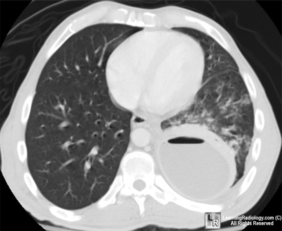
Streptococcus Anginosus Empyema

Today we discussed a patient with a chronic empyema ultimately proven to be secondary to streptococcus anginosus.

Please follow the link to a recent review on parapneumonic effusions and empyema.

Some take-home points about S.anginosus and empyema:

1) The Streptococcus milleri group of organisms is made up of S. intermedius, S. anginosus and S. constellatus which are considered a sub-group of Viridans streptococci. These gram positive cocci are normal flora in the respiratory and GI tract and are known to cause abscesses in the brain, liver, lungs and oropharynx.

2) S.milleri are generally susceptible to beta-lactam antibiotics.

3) In general, lung abscesses related to S. milleri are thought to occur from aspiration of oropharyngeal contents.

4) Don't forget to look for the caramel of butterscotch smell associated with S. anginosus infections! Actually no, please don't.

5) The management includes prompt drainage of any collections and IV antibiotic therapy, usually with a beta-lactam. Duration of treatment is dependent on symptoms, but often a minimum 4 week duration of IV antibiotics is required. Specific antibiotic selection should be based on sensitivity testing.

6) Regarding complicated parapneumonic effusions and empyema:

a) These effusions typically occur in the context of pneumonia and represent a spectrum from an uncomplicated parapneumonic (UPPE) effusion to a complicated parapneumonic effusion (CPPE)to an empyema.

b) The signs and symptoms of an empyema are often indistinguishable from a typical pneumonia. All patients with pneumonia should be assessed for a pleural effusion.

c) In the setting of pneumonia with a pleural effusion, the phrase "never let the sun set on a parapneumonic effusion" is used to emphasize the importance of prompt investigation and drainage if necessary. Effusions greater than 10mm on the lateral decubitus CXR should be sampled.

d) More chronic presentations of an empyema can occur with less virulent organisms such as S.anginosus (as in our patient).

e) In general, a PPE should be drained if the pH of the fluid is less than 7.2, there is a positive gram stain/culture, a glucose less than 40-60 (U.S. units), an LDH greater than 1000 (U.S. units) or the effusion is greater than 50% of the volume of the hemi-thorax.

f) Drainage of a CPPE or empyema typically involves a large bore chest tube +/- adjunctive therapies such as fibrinolytics, VATS or decortication.


Monday, November 22, 2010

Antibiotics and Pneumonia


Today we talked today about a patient with community acquired pneumonia (CAP) and the appropriate antibiotics and investigations for CAP.


Here's a link to the 2007 IDSA/ATS CAP Guidelines (scroll down to CAP). The BTS also released guidelines in 2009.

In brief:


1) While it seems obvious, an infiltrate on CXR (in the right clinical context) is required for the diagnosis of pneumonia.

2) Generally, all patients with pneumonia admitted to hospital should have blood and sputum cultures drawn.

3) In patients with severe pneumonia, urinary antigens for Legionella pneumophila and Streptococcus pneumoniae should be sent.

4) Options for outpatient treatment:
a) If previously healthy, can use a macrolide or doxycyline.
b) In patients with comorbidities or recent Abx use, give a respiratory FQ or a (beta- lactam + a macrolide).
c) Remember, if the presence of macrolide resistant S.pneumo is greater than 25% in your practice area, macrolides should be avoided.

5) Inpatient, non-ICU treatment:
a) (Respiratory FQ) or a (beta-lactam + a macrolide).

6) Inpatient, ICU treatment:
a) A beta-lactam plus either a macrolide or a FQ (better evidence for FQ over macrolide).
b) If concerned re: pseudomonas, give an anti-pseudomonal beta-lactam that has anti-pneumococcal activity (PipTazo, Cefepime, Imipenem or Meropenem) plus either (Cipro of Levo) or (an aminoglycoside + azithromycin) or (an aminoglycoside + an anti-pseudomonal FQ).
c) Add Vano or Linezolid if MRSA risk factors present.

7) The beta-lactam in the outpatient and in-patient not ICU treatment arms can be high dose amoxicillin/ampicillin.

Fever of Unknown Origin


Last week Friday at Rheumatology Morning Report we discussed a patient with a fever of unknown origin (FUO).

This is a challenging Internal Medicine presentation with limited high quality evidence and a wide differential diagnosis.

A number of reviews have been published in recent years offering varied approaches to making a diagnosis. Take a look at our own Toronto approach published by Drs. Mourad, Detsky and Palda in the Archives of Internal Medicine. Two other reviews include one from AFP and another from Postgraduate Medicine.

A few points about FUO:

1) The classical definition of FUO is a temperature greater than 38.3C on multiple occasions over a period of 3 weeks with no diagnosis found after 1 week of inpatient investigations.

While this definition is likely outdated in the era of expanded outpatient management, the bottom line is that there must be a documented fever for a prolonged period of time. This is done in an attempt to exclude any transient viral infections. One modern definition suggests that the fever should persist for more than 3 weeks despite 3 outpatient visits and/or 3 days of in-hospital investigations to be considered an FUO.

The classic definition and approach to FUO typically excludes patients with HIV, immunosuppression or neutropenia.

2) The incidence of FUO has been estimated in one series to be less than 3% of admitted medical patients. The frequency of FUO continues to decline with the advent of increasingly sophisticated investigative modalities (cultures, serology, radiology).

3) The etiologies of FUO can be grouped into 5 categories: infection (16%), inflammation (22%), malignancy (7%), miscellaneous (4%) and undetermined (51%).

4) As always, the investigation should start with a focused history and physical examination. Remember to pay special attention to recent travel, medication use (including anti-pyretics), possible immunosuppression, the pattern of fever and any localizing symptoms.

5) A number of initial investigations have been suggested. One approach is to ensure the patient has a minimum set of investigations before proceeding down a FUO algorithm.

These might include:
i) CBC and differential
ii) Blood film
iii) 3 set of blood cultures (separated in space and time)
iv) Lytes, Cr, HCO3, LDH, liver enzymes and function tests
v) Urinalysis and culture
vi) CXR

From here if a source of fever has not be elucidated, further minimum investigations might include:

i) ANA and RF
ii) HIV testing
iii) CMV IgM or heterophile testing (if suggested clinically)
iv) Hepatitis serologies (if liver enzymes elevated)
v) ESR or CRP
vi) One article suggests Q-fever serology (if suspected clinically)
vii) One article suggests an SPEP (if MM suspected clinically)
viii) CK
ix) A TB skin test has been suggested in some articles

If this second set of investigations fails to yield of a diagnosis, all non-essential medications should be discontinued had imaging should be pursued:

i) CT chest/abdomen - assess for lymphadenopathy or collections
ii) One article suggests a technetium scan in addition to the above CTs.

Following these investigations, if no etiology is apparent, then further testing should increasingly be focused towards the most likely diagnosis. This may include:

i) Echocardiography - assess for valvular vegetations
ii) Leg dopplers - assess for a silent DVT
iii) Temporal artery biopsy - in patients greater than 50 years old to assess for TA
iv) Liver biopsy - if clinically deteriorating
v) Bone marrow biopsy - if abnormalities are seen in the CBC and blood film

6) Therapeutic Trials
In general, therapeutic trials of antibiotics or corticosteroids are discouraged unless a thorough hunt for an etiology has been completed. This is out of concern of exacerbating an undiagnosed infection (steroids), sterilizing cultures (antibiotics) or suppressing a fever while the etiology remains.

7) Prognosis
If no diagnosis is found after an extensive evaluation, the prognosis of these patients is generally good.

Wednesday, November 17, 2010

Pulmonary Embolism and IVC Filters


Today we discussed the diagnosis and management of a pulmonary embolism (PE) and the role of Inferior Vena Cava (IVC) filters.

The role of IVC filters in PE remains controversial and there is significant variability in clinical practice. Guidelines from the ACCP exist, however recent evidence suggests that up 25% of IVC filter insertions are done for indications that do not follow the guidelines.

As mentioned today, IVC filters can be associated with catastrophic consequences in the event of filter fracture and embolism as discussed in this 2010 study.

Here's an excellent recent review on acute PE from the NEJM this year and a link to the PIOPED II Trial we discussed today.

A few points on the diagnosis and management of a PE with a focus on IVC filters:

1) The diagnosis or exclusion of a PE is heavily influenced by the pre-test probability before objective testing begins. The Well's criteria is a well validated scoring system to estimate the pre-test probability of a PE:

i) Clinical symptoms of a DVT - (3 points)
ii) Other dx less likely than PE - (3 points)
iii) HR greater than 100 - (1.5 points)
iv) Immobilization for more than 3 days or
surgery within the past 4 weeks - (1.5 points)
v) Previous DVT/PE - (1.5 points)
vi) Hemoptysis - (1 point)
vii) Malignancy - (1 point)

A score of greater than 4 is considered "PE likely" whereas a score of 4 or less is considered "PE unlikely". A reasonable approach is to pursue CTA in the "PE likely" group and consider d-dimer (ELISA) testing in the "PE unlikely" group. One caveat is that most of these studies were done in outpatients and the approach of using d-dimer testing may not be generalizable to admitted GIM patients. Often this means going directly to CTA.

Remember, PIOPED II demonstrated that if the results of CTA are discordant with your clinical assessment (i.e. CTA negative in a high pre-test probability patient or positive in a low risk patient), this should promote additional testing.

2) After the diagnosis is made, anticoagulation should be initiated with heparin first (preferably LMWH over IV heparin) or Fondaparinux. The patient can then be bridged onto warfarin for a minimum of 3-6 months of anticoagulation. The exception to this is in patients with an active malignancy or receiving chemo where the CLOT Trial showed that anticoagulation with LMWH is superior to warfarin in preventing recurrent VTE and may reduce mortality in patients with solid tumors (sub-group analysis).

3) Consider thrombolysis in patients with hemodynamic instability from an acute PE.

4) The only indication for IVC filter insertion recommended in the 2008 ACCP Guidelines is for patients with a PE and a contraindication to anticoagulation. Furthermore, if an IVC filter is inserted, anticoagulation should be re-instituted as soon as is deemed safe.

5) Some authorities suggest inserting an IVC filter in the context of a recurrent pulmonary embolism while on anti-coagulation.

6) Controversy exists regarding the role of IVC filter placement in patients with poor cardiopulmonary reserve or a large clot burden.

7) "Prophylactic" IVC filters should be avoided.

8) The largest trials to date have shown that in hemodynamically stable patients, IVC filters reduce the incidence of recurrent symptomatic PEs, but do not change mortality and cause an increased rate of DVTs (when left in). The benefits may be greater in hemodynamically unstable patients.

9) Most IVC filters are retrievable! Talk to Interventional Radiology and arrange to have them removed. Do not leave your patient with a long term IVC filter unless they still need it!

Tuesday, November 16, 2010

Portal Vein Thrombosis

Today we discussed a patient with a portal vein thrombosis (PVT) in the setting of acute cholecystitis. Following the management of acute cholecystitis, the discussion turned to the management of a PVT.

Here's an excellent review article on an approach to managing a PVT.

A few things to keep in mind:

1) The causes of a PVT include inflammatory, malignant, infectious and hypercoaguable states, however the most common cause is cirrhosis (25%).

2) An incidentally found PVT should trigger a search for cirrhosis, a hypercoaguable state or occult malignancy (including a myeloproliferative disorder).

3) An acute PVT is usually asymptomatic.

4) The rationale for treating a PVT is to prevent clot extension and compromise of intestinal perfusion and to prevent portal HTN.

5) It's estimated that 85-90% of patients with a chronic PVT thrombosis have esophageal varices.

6) There is very limited evidence to inform the decision to anticoagulate or not. Management is often extrapolated from the lower extremity DVT literature.

7) In the context of cirrhosis, anticoagulation is generally NOT recommended due to the increased risk of bleeding.

8) In a chronic PVT with portal HTN and NO cirrhosis, it may be reasonable to anticoagulate if there is a history of a hypercoaguable state or previous thrombosis. Otherwise, the risk of bleeding in the context of portal HTN (varices) may outweigh the benefits.

10) In an acute PVT and NO cirrhosis, it is reasonable to investigate for and treat any underlying etiology (including a hypercoaguable state) and consider anticoagulating for a minimum of 3-6 months or longer if there is a persistent risk factor.

11) If anticoagulating, the same medications (warfarin, LMWH, IV heparin) and targets (i.e. INR 2-3) can be used as for lower extremity DVTs.

12) The management of a PVT is highly variable, clinician dependent and must be tailored to the individual patient and clinical scenario.

Monday, November 15, 2010

ASA Overdose


This morning we talked about the approach to an aspirin (ASA) overdose. This is an important topic to review since we don't see it very frequently on GIM yet its management requires time sensitive interventions to prevent morbidity and mortality.

Here's a flow chart from the BMJ. If you have access, here's a review article from Postgraduate Medicine. The Merck Manual also has a short review as does Harrison's. The Uptodate article on "Aspirin Poisoning in Adults" is also a nice summary.

We talked about urine alkalinization quite a bit today. Here's a link to a position paper published in the Journal of Toxicology that supports urinary alkalinization.

Some key points:

1) Salicylates are found not only in aspirin (acetylsalicylic acid), but also in a number of OTC/herbal products including Bismuth subsalicylate (Pepto Bismol), salicylic acid and Oil of Wintergreen.

2) In general (watch your units):
i) Therapeutic Range: 0.7-2.2mmol/L
ii) Toxic Range: >greater than 2.9mmol/L
iii) Consider dialysis when level is greater than 7.2mmol/L

3) In usual doses, aspirin is rapidly absorbed and reaches a peak concentration in 1 hour. However, in the setting of an overdose, this can be significantly delayed (5-6 hours from ingestion) due to slowed gastric emptying or enteric coated/slow-release preparations.

4) Pathophysiology of Toxicity:
i) Uncoupling of oxidative phosphorylation at the cellular level
ii) Stimulation of the respiratory centre in the medulla
iii) Metabolic acidosis (primary from salicyltes, secondary from mitochondrial poisoning)
iv) Central hypoglycemia despite normal peripheral blood glucose levels

5) Signs and Symptoms of overdose: nausea, vomiting, tinnitus, hyperventilation. More severe symptoms include: fever, decreased LOC, non-cardiogenic pulmonary edema, rhabdomyolysis, hypotension, seizures and AKI.

6) When suspecting any overdose, always check a Tylenol and ASA level.

7) Important investigations: ASA level (check q2h until two values show decreasing levels), ABG (or venous blood gas), CBC, lytes, creatinine, liver enzymes, INR, plasma glucose, extended electrolytes, urinalysis, urine pH and CXR. The blood work and urine tests should be repeated frequently during the initial period of management.

8) ASA intoxication is one of the prototypical causes of a mixed repiratory alkalosis with an anion gap metabolic acidosis (although an isolated anion gap metabolic acidosis can be seen).

9) Remember the common causes of an anion gap metabolic acidosis: methanol, uremia, diabetic/starvation/alcoholic ketoacidosis, lactic acidosis, ethylene glycol, salicylates.

10) Management:

i) Stabilize ABCs.

ii) If tolerated, activated charcoal should be given (1g/Kg up to 50g) and can be repeated q4h x 2 after the first dose. This works best if given within 1 hour of toxic ingestion. Contraindicated if level of consciousness is depressed unless the patient is intubated.

iii) Supplemental glucose should be given in patients with altered LOC due to the risk of neuroglycopenia despite normal peripheral blood glucose.

iv) Potassium must be aggressively replaced. Hypokalemia can impair urine alkalinization, and any initial hypokalemia will be exacerabted by alkalinization.

v) Urine alkalinization: This is a critical step. Here's a position paper published in the Journal of Toxicology on the topic.

Salicylic acid is a weak acid. The non-ionic form can diffuse across membranes, while the ionic form cannot. Increasing the serum pH will result in diffucion of the non-ionic form out of cells and trap it in the blood/urine in the ionic form.

Pre-existing alkalosis is not a contraindication to urinary alkalinization.

A rise in the urine pH of 1 unit can theoretically increase salicylate excretion by a factor of 10.

Start with a bolus of 1-2mEq/Kg of NaHCO3 followed by an infusion of 3amps of NaHCO3 mixed in 850cc D5W (NOT normal saline) run at 1.5-2 times maintenance. Urine pH should be checked frequently with a target of > 7.5 - 8. Failure to achieve this within 1-2 hours may be an indication for dialysis.

11) Avoid acetazolamide and any medication that depresses respiratory drive.

12) Indications for dialysis:
i) Renal failure impairing salicylate clearance
ii) Inability to alkalinize the urine
iii) Fluid overload limiting sodium bicarbonate treatment
iv) Plasma salicylate level greater than 7.2 mmol/L
v) Coma
vi) Failure of medical management

13) Always call your local poison centre for advice when managing an overdose.

Friday, November 12, 2010

Hemorrhage

Yesterday we talked about a patient presenting with a Hg of 42 and an MCV of 50 with no signs of bleeding. During our discussion, a number of topics related to anemia, hemorrhage and transfusions came up.

We talked briefly about the TRICC trial published in the NEJM. This study looked at 838 ICU patients with a Hg of less than 90 and randomized them to a liberal (transfuse if Hg less than 100) vs. a conservative (transfuse if Hg less than 70) transfusion strategy.

The study showed that at 30 days there was no between arm difference in mortality but there was reduced in-hospital mortality in the restrictive transfusion arm. In sub-group analysis, this arm also demonstrated reduced mortality in younger patients (less than 55 years old) and less acutely ill patients (APACHE-II score less than 20). No difference in outcome was seen between the arms in patients with an ACS.

TRICC Bottom Line:

1) These were ICU patients so be cautious with generalizing to GIM patients, but we generally follow the results from TRICC.

2) Consider a RBC transfusion in patients with a Hg of less than 70, especially if symptomatic. This is not a hard and fast rule - you may not transfuse a patient with a Hg of 65 who is well and asymptomatic, especially if it is chronic.

3) Consider not transfusing RBCs in a patient with a Hg greater than 100. However, it may be reasonable to aim for a higher Hg (i.e. greater than 100) in patients with an acute coronary syndrome.

4) A significant gray zone exists inbetween 70 and 100.

Sources of Blood Loss:

1) When presented with a very low Hg, you need to rule out immediate life theatening causes right away - bleeding and hemolysis.

2) A reasonable initial screen for hemolysis includes an LDH, haptolgobin, blood film and bilirubin. The combination of an elevated LDH and low haptoglobin are 90% specific and 92% sensitive for hemolysis.

3) Looks for obvious sources of bleeding: GI tract (BRBPR, melena, hematemesis), GU tract, trauma (laceration, hemo-thorax/peritoneum, etc.) or Gyne.

4) If no obvious source, consider hemolysis and/or a retroperitoneal bleed.

5) Large hemodynamically significant blood loss will not occur from an intracranial hemorrhage due to the limited space in which there is to bleed. Additionally, in a pulmonary hemorrhage, hypoxia will be a greater threat to life before anemia is.

A few points on microcytic anemias:

1) We talked briefly about differentiating iron deficiency anemia from thalassemia. Thank you to Lauren for pointing out that the RDW may be useful in this case. In thalassemia, the RDW is generally low or normal whereas in iron deficiency anemia it may be elevated as the bone marrow pushes out RBC precursors.

2) In a patient over the age of 50 with iron deficiency anemia, remember to look for occult bleeding from a GI malignancy.

3) A ferritin of less than 10-15 is virtually diagnostic of iron deficiency anemia (99% specific, 59% sensitive). Using a value of 41 increases the sensitivity to 98% and the specificity to 99%. The challenge is that ferritin is an acute phase reactant and is increased in inflammation. One possible approach is to divide the ferritin value by three in these patients, if it's less than 20, this is highly suggestive of co-existing iron deficiency anemia.

4) Don't forget celiac disease as a not uncommon cause of iron deficiency anemia.

5) Many other tests including iron saturation, TIBC, Hg electrophoresis, reticulocyte count, soluble transferrin receptor and blood film examination can help in the assessment of a microcytic anemia.


Wednesday, November 10, 2010

Atrial Fibrillation

Today we talked about atrial fibrillation in a patient with a history of rheumatic fever. On GIM, atrial fibrillation is a diagnosis we see very commonly.

One of the best atrial fibrillation resources is the 2006 ACC/AHA Guidelines which you can find here. As a side note, the ACC/AHA have guidelines on a huge variety of cardiovascular topics that provide an evidence base for cardiac decision making on the wards. Here's the list.

Briefly about atrial fibrillation:

1. Afib is a very common arrhythmia. Seen in ~10% of patients greater than 85 years old.

2. While afib can cause distressing symptoms (palpitations, SOB, etc.), the biggest concern is thromboembolism and the impact of afib on cardiac function.

3. Generally we classify afib as:
a) paroxysmal (less than 7 days duration)
b) persistent (lasting greater than 7 days and terminating spontaneously or with cardioversion)
c) permanent (afib lasting greater than 1year and failed cardioversion/not attempted).

Regardless of the classification,
they all portend the same yearly risk of stroke.

A fourth classification is "lone atrial fibrillation" which applies to atrial fibrillation in a patient with no structural heart disease (and often less than 60 years old). These patients are generally felt to be at a low risk of stroke.

4. Most common causes of afib: HTN, valvular disease, hyperthyroidism, heart failure and alcohol.

Usually afib is not associated with stable coronary artery disease unless it results in an MI or heart failure. Afib can also be seen in the context of surgery or a pulmonary embolism.

5. Minimum set of investigations for new onset atrial fibrillation: ECG (confirm diagnosis, assess for other complicating disease), CBC, lytes, TSH and 2D Echo (to assess for structural heart disease).

6. Management consists of rate vs. rhythm control and thromboembolism risk reduction.

a) Rate vs. rhythm control:

Often a trial of cardioversion is given for the first episode of atrial fibrillation. In the long term, most patients with recurrent atrial fibrillation are managed with rate control rather than rhythm control (unless they have significant symptoms or cardiovascular compromise when in afib).

The AFFIRM Trial demonstrated that there was no survival or stroke risk advantage to a rhythm control strategy and that rhythm control more frequently resulted in medication side effects and hospitalization.

Rate control is usually accomplished with B-blockers, non-dihydropyridine calcium channel blockers (Dilt, Verapamil) or Digoxin. The guidelines suggest a HR target of less than 80bpm at rest and less than 110bpm with exercise. However, the recently published RACE-II Trial suggests that strict rate control may confer no additional benefit over lenient rate control.
b) Thromboembolism risk reduction:

The yearly risk of stroke depends on a variety of factors. Several risk scores exist, but one of the easiest to use is the CHADS2 Score. This only applies for patients with non-valvular atrial fibrillation:

Chronic Heart Failure - 1 point
HTN - 1 point
Age greater than 75 - 1 point
Diabetes - 1 point
Stroke/TIA - 2 points

If the CHADS2 score is 0 then no therapy or ASA is reasonable. With a CHADS2 score of 1 then ASA or Warfarin (INR 2-3) is acceptable and with a CHADS2 score greater than 1 warfarin is recommended.

The reason for this is that the benefit you obtain from ASA or warfarin is determined by your yearly risk of stroke. ASA provides a RRR of ~25% while warfarin gives a RRR of ~66%. The risk of a significant bleeding complication from ASA is small, while with warfarin is ~1%/year (depending on the patient).

Remember, for patients with valvular atrial fibrillation, their stroke risk is much higher (as in our patient this morning) so they should be managed with warfarin.

Interestingly, the recently published RE-LY trial showed that Dabigatran, a direct thrombin inhibitor that does not require monitoring may be superior to warfarin in terms of stroke prevention or bleeding risk (depending on the dose). This could revolutionize the way atrial fibrillation in managed.


Tuesday, November 9, 2010

Acute Hepatitis

Today we discussed a case of acute hepatitis NYD with moderate-severe liver enzyme elevation. This is not an uncommon referral to Internal Medicine.

Here's a very useful review from CMAJ in 2005 on elevated liver enzymes.

Some points about marked liver enzyme elevation (>5x the upper limit of normal):

1. Remember that the "liver function tests" are bilirubin, INR, albumin +/- glucose and can reflect the synthetic function of the liver. Conversely, the "liver enzymes" - AST, ALT, ALP and GGT are NOT liver function tests as they offer no information on the liver's synthetic function but merely are a marker of hepatocyte integrity and/or cholestasis.

2. The differential diagnosis of moderate to severe transaminitis includes: viral hepatitis, toxic hepatitis, ischemic hepatitis, severe obstruction, autoimmune and alcoholic hepatitis.

3. Decide what the predominant pattern is to help you guide investigations:
a) Hepatic (increased AST, ALT > ALP)
b) Cholestatic (increased ALP > AST, ALT)

4. How elevated are the enzymes? 5x vs. 5-10x vs. >10x of normal? What is the trend? Is there synthetic dysfunction (increased INR, bilirubin, decreased albumin)? Typically liver enzyme values at this level represent acute liver injury.

5. In acute viral hepatitis, the liver enzymes often peak before the bilirubin and the patient may have non-specific symptoms such as fatigue, arthralgias and a low grade fever (as in our patient) .

6. Consider ischemic hepatitis in the right clinical context and an ALT/LDH ratio of less than 1.

7. Hepatitis screen: Hep A IgM Ab, Hep B core Ab (IgM), Hep B surface Ag and Ab, Hep C Ab.

8. Always ask about Tylenol use, herbal and over the counter medications, periods of illness (hypotension), autoimmune symptoms and alcohol.

9. Some form of abdominal imaging (usually ultrasound) is often appropriate to assess for evidence of hepatitis and/or obstruction.

10. Once the common causes of moderate to severe transaminitis have been ruled out, consider testing for auto-immune hepatitis and other viral causes such as EBV and CMV.


Monday, November 8, 2010

Knee Pain

Yesterday we talked about the diagnostic dilemma of a painful knee that ultimately was presumed to be secondary to osteoarthritis.

This a good opportunity to review the approach to an acute monoarthritis and to osteoarthritis.

Let's start with some Canadian content. Here's a recent review published in the CMAJ looking at the management of a patient with an acute monoarthritis.

The NEJM published a very thorough review article on OA of the knee in 2006.

There are many causes of knee pain outside of a monoarthritis or osteoarthritis, many of which we do not see very often as internists (i.e. orthopedic problems in younger patients).


To summarize, here are some key points on evaluating a patient with acute knee pain:

1. Determine if there is a recent history of trauma. This raises the pre-test probability of a fracture, hemarthrosis, meniscal tear or ligamentous injury.

2. Decide whether this is an intra-articular, peri-articular or referred process (there is significant overlap in some of these signs/symptoms):

a) Intra-articular: pain localized to the knee without radiation, knee effusion, mechanical symptoms (clicking, locking, giving way), abnormal structural physical exam, reduced range of motion.

b) Peri-articular: focal area of pain or abnormalities on exam, pain only during parts of the range of motion, +/- mechanical symptoms.

c) Referred pain: vague location of pain, back or hip pain, pain not exacerbated by knee movement, lack of an effusion, normal knee exam.

3. If the pain is articular, you need to decide if it is inflammatory (i.e. arthritis - pain, redness, swelling, warmth) or non-inflammatory (structural). Any acute effusion should be drained and analyzed.

4. Remember if an effusion is present -> rule out INFECTION. Missing a septic joint can have catastrophic consequences for the joint, and very quickly.

5. Arthrocentesis fluid should be sent for cell count and differential, gram stain, C&S, microscopy for crystals at a minimum.

6. Imaging: Start with x-rays. If there is any history of trauma, lack of localizing symptoms to the knee or a question of pain origin, image the ipsilateral joints above and below the knee.

7. Consider MRI as the next imaging test of choice after x-ray if no diagnosis can be found or if a soft tissue etiology is being considered.

8. An effusion can be present in an acute exacerbation of osteoarthritis.


Friday, November 5, 2010

Renal Cell Carcinoma

Today we talked about renal cell carcinoma presenting with anorexia, abdominal distension, hematuria, ascites and renal failure.

Here are links to two review articles, the first is from the Lancet in 2009 and the second is from NEJM in 2005.

Some key points about RCC:

1) RCC is the 7th most common cancer in men and the 9th most common in women. The peak incidence is in the 6th and 7th decades of life (mean age of 60). Men are affected twice as frequently as women.

2) Risk factors include: HTN, obesity, smoking, acquired cystic diseases of the kidney, ESRD and certain inherited conditions (e.g. von Hippel-Lindau). Regardless, most patients with RCC do not have an identifiable risk factor. Fruit and vegetable consumption may be protective!

3) This tumor was classically known as "The Internist's Tumor" due to its varied presentation, however with the widespread use of abdominal imaging and incidental diagnosis, some argue that RCC is now "The Radiologist's Tumor".

4) Signs and Symptoms at Presentation:
a) Local: The classic triad of abdominal pain, a palpable mass and hematuria is uncommonly seen today.
b) Systemic: Anorexia, fatigue, wasting and a variety of paraneoplastic phenomena including hypercalcemia (PTHrp), HTN (renin) and erythrocytosis (EPO).

5) Any patient over the age of 40 with hematuria should be assessed for RCC since prognosis is determined by stage.

6) Imaging: CT abdomen is the investigation of choice. However, abdominal U/S while less sensitive can help differentiate a simple cyst from a more complex structure. MRI can be useful if the U/S is non-diagnostic or contrast dye is contraindicated.

7) If a solid renal mass is identified with radiologic features consistent with RCC, biopsy is usually not performed due to low specificty and concern with seeding the peritoneum. Good surgical candidates often go directly for a partial or radical nephrectomy.

8) Surgical resection is the cornerstone of therapy as chemotherapy is minimally effective. Surgery can be curative in the majority of patients with Stage I, II and III disease. Resection of the primary tumor is recommended even in the setting of metastatic disease (in patients with good functional status).

9) Significant interest is currently focused on biologic agents including VEGF and mTOR inhibitors given the limited benefit and significant toxicity of Interleukin-2 immunotherapy.

Thursday, November 4, 2010

The Art of Pimping

Hi folks,

Usually we keep things pretty formal and academic here on Horses and Zebras, but today we're going to talk about something different - the art of pimping.

For those unfamiliar with the term as it applies to Medicine, this is when a staff "poses a series of very difficult questions to an intern or student." Often this is done to further learning or highlight gaps in knowledge, but at times it can make the learner feel self-conscious or even uncomfortable.

We've all experienced this and part of going through your training is knowing how to handle it.

Check out the very humorous, but all too true article that started it all. Some excellent defensive tips are offered.

Our own Dr. Detsky furthered the discussion on this topic with his article from JAMA in 2009.

In all seriousness, "pimping" will continue to be a part of medical education for the foreseeable future. Here are some things (from one trainee to another) that have helped get me though the 10,000 times I've been put on the spot:

1) Remember that your staff is trying to help you learn, not hurt your feelings. You will never forget something you got wrong when pimped (i.e. the Toronto clinical prediction rule for aortic stenosis).

2) It's OK to stop, take a deep breath and think about the answer rather than blurting out the first thing that comes to mind.

3) If you don't know the answer or are never going to remember it, just be honest and say, "I don't know." This is better than squirming for 5 minutes during which time it'll be obvious you didn't know the answer.

4) If you think you know the answer, then answer with confidence. Not arrogance, but confidence. If you're right, you'll get credit for being right! If you're wrong - you were going to be wrong anyway, so you've lost nothing. However, if you're right but you answered in a sheepish, high pitched and uncertain voice, you'll only get credit for guessing.

Finally, when it's your turn to be the teacher, remember what it felt like to get "pimped".

Wednesday, October 27, 2010

Chronic Diarrhea


Today we talked about a patient with chronic diarrhea.

Here is a useful review article from Gastroenterology in 2004.

Guidelines from the American Gastroenterology Association were published in 1999.

Working up chronic diarrhea can be challening as the differential diagnosis is quite broad, clinical trials are lacking and expert opinion is varied.

Some key points:

1) Chronic diarrhea is not well defined. A reasonable definition might be:

  • Decrease in stool consistency ("stool that takes the shape of the vessel it is in")
  • >3 stools and/or >200g of stool per day
  • >4 weeks duration

2) Have an organized approach to the types of chronic diarrhea (which leads to the differential). The guidelines suggest watery vs. inflammatory vs. fatty. Another classification scheme includes secretory vs. malabsorption/osmotic vs. inflammatory.

3) Use the history and physical exam to direct your initial set of investigations. Make sure to take a good medication and travel history!

4) Reasonable initial tests beyond the routine:

  • Stool osmotic gap [290 - 2(stool Na + stool K)] and pH
  • FOBT
  • TSH
  • anti-TTG and IgA level (if risk factors for celiac)
  • Stool C&S, O&P (repeat over several days) and C.diff
  • Fecal WBCs
  • Response to fasting
  • +/- 72 hour fecal fat testing (difficult to get done)
  • +/- Colonoscopy or Sigmoidoscopy
  • +/- Abdominal imaging

5) Stool osmotic gap (see above):

  • a value less than 50 suggests secretory diarrhea (i.e. lots of secreted electrolytes in the lumen)
  • a value greater than 125 suggests osmotic diarrhea (i.e. lots of unmeasured osmoles in lumen)
  • values in between are indeterminate

6) Testing for pancreatic insufficiency, bacterial overgrowth, hormone levels (gastrin, calcitonin, VIP, carcinoid), mucosal abnormalities, etc. can be pursued as appropriate following the initial set of investigations.

7) Some authors suggest that more than 80% of chronic diarrhea cases have a treatable etiology.

Tuesday, October 26, 2010

Hemoptysis

Hey folks,

Horses and Zebras is back after a brief absence!

Today we talked about a patient with new onset hemoptysis and an otherwise normal physical exam, screening blood work and CXR.

Here's a short and focussed review article from AFP on hemoptysis. It includes a very easy to follow algorithm and a good differential diagnosis.

A couple of key take-home points:

1) Always start with the ABCs. While we worry about dropping hemoglobin and hypotension, remember that one of the biggest risks in patients with hemoptysis is airway compromise and hypoxia.

2) Decide if this is actually hemoptysis! Blood from the GI tract or above the vocal cords (pseudohemoptysis) can mimic hemoptysis.

3) Infection and cancer are the most common diagnoses.

4) Evaluate for massive hemoptysis (>200-600cc/24 hours). This may suggest a diagnosis but also implies a much higher risk of airway compromise and need for aggressive airway management.

5) Reverse the reversible - correct coagulopathies, thrombocytpenia, etc.

6) After routine bloodwork - get a CXR. Go on to bronchoscopy or a high resolution CT scan of the chest in patients with massive hemoptysis, persistent bleeding, CXR findings or risk factors for lung cancer.

7) Call for help sooner rather than later - ICU, respirology, thoracic surgery - if the bleeding is not resolving or is getting worse. Specialized airway skills may be needed.

Friday, September 17, 2010

Multiple Myeloma


Today we discussed a classic case of Multiple Myeloma with hypercalcemia, renal failure, anemia, and bone pain secondary to lytic bone lesions... these features make up the mnemonic CRAB: hyperCalcemia, Renal failure, Anemia, Bone pain.

Here is a link to a previous Horses & Zebras blog summarizing the Clinical Features of Multiple Myeloma, as well as some links to articles reviewing investigation and management of hypercalcemia and current management options for multiple myeloma.

Management of hypercalcemia +/- acute renal failure is often the primary problem that needs to be managed on admission to hospital. Hypercalcemia can be considered to be mild (2.6 - 2.9 mmol/L), moderate (3.0 - 3.4 mmol/L), and severe (greater than 3.5 mmol/L); however, there are no official cut-off points for hypercalcemia. As we discussed this morning, ~40% of calcium is bound to albumin and thus low albumin states will lower the measured calcium. This can be corrected roughly by adding 0.2 mmol/L to a calcium measurement for every drop in albumin of ~10.

Treatment of Hypercalcemia

1. First line treatment for hypercalcemia is fluid resuscitation, as the majority of patients will be volume contracted. Rapid, aggressive resuscitation is the goal, monitoring for any potential volume overload/heart failure in susceptible patients.

2. Administration of an IV bisphosphonate is also considered appropriate in severe hypercalcemia. Pamidronate (60 - 90 mg) is the usual choice, given once over 2-4 hours, and most often normalizing calcium within 5-7 days.

3. For more immediate calcium-lowering calcitonin can be considered; however, it is not uncommon for patients to develop tachyphylaxis and thus its use is limited.

4. Alternatively, and often more effective in the setting of lymphoma or multiple myeloma induced hypercalcemia, glucocorticoids such as prednisone (1 mg/kg daily) can be considered.

5. Management of hypercalcemia always includes investigation into the underlying cause, and treatment of such problem otherwise the hypercalcemia will recur.

Friday, September 10, 2010

Guillain-Barre Syndrome

This week we explored 2 different (potential) cases of Guillain-Barre Syndrome, or GBS. First during Physical Examination Rounds and then again in Morning Report.

The most common form of GBS is an idiopathic acute inflammatory demyelinating polyneuropathy (AIDP), and in fact GBS is often synonymous for AIDP. It is an autoimmune process directed against Schwann cell membranes. There are 5 other variants of GBS, including the Miller Fisher Variant which often presents with the triad of ophthalmoplegia, ataxia, and areflexia.

GBS occurs anywhere from 1 - 2 per 100,000 per year. It is often (~2/3 of cases) preceded by either a respiratory or GI infection, most commonly Campylobacter. GBS has also been reported after vaccinations and to be associated with systemic illnesses, such as SLE, Hodgkin's Lymphoma, Sarcoidosis, etc.

Patients with GBS often present with parasthesias in their limbs, followed soon after by ascending weakness. This progresses over days to weeks. GBS can also be associated with autonomic dsyfunction such as orthostasis, urinary retention, constipation, and tachycardia.

Physical examination most often reveals symmetrical motor weakness in an ascending pattern, with absent reflexes, flaccid tone, and a normal sensory exam.

Patients with GBS need to be monitored closely for respiratory failure due to involvement of the diaphragm. A good rule of thumb for consideration of intubation is the 20/30/40 rule:
  • 20: Vital Capacity less than 20 cc/kg
  • 30: Maximum Inspiratory Pressure less than 30 cm H2O
  • 40: Maximum Expiratory Pressure less than 40 cm H2O

Treatment for GBS is most often supportive, and may require an ICU setting for ventilatory support, hemodynamic support, nutritional support, and pain control. If diagnosed within the first 4 weeks then IVIG or plasmapheresis can be considered. A recent Cochrane review suggests that IVIG treatment has similar outcomes to plasmapheresis in the setting of early (ie: within 2 weeks) diagnosed GBS.

Outcomes for patients with GBS are quite good with ~80% achieving full recovery within a few months to a year; however, minor deficits often persist. 5-10% will have permament disabling deficits, and another 2-3% will die as a result of their GBS. Approximately 5-10% of patients may develop one or more relapses of their GBS, leading to a diagnosis of Chronic Inflammatory Demyelinating Polyneuropathy (CIPD).

Here is a very thorough review of GBS from 2005, in The Lancet.

Friday, September 3, 2010

Pulmonary Hypertension


Today we discussed a case of newly diagnosed Pulmonary Hypertension in a young woman.

Pulmonary Hypertension (PH) is defined as a Pulmonary Arterial Pressure over 25 mmHg at rest (normal 12-16 mmHg).

Patients often present with months or years of gradually progressive symptoms, such as shortness of breath, fatigue, cough, angina, peripheral edema, and rarely hemoptysis.

Cardiovascular examination often reveals a loud, palpable P2, a split S2, and an RV heave.

There are multiple causes of PH, but all result in increased pressures within the pulmonary vasculature, either due to vasoconstriction of the blood vessels, obstruction by way of a mass or pulmonary emboli, or fibrosis within the lung parenchyma related to systemic illnesses. In order to overcome this increased pulmonary pressure, the heart then is required to create elevated right-sided pressures in the heart. In turn, this leads to RV hypertrophy and potentially RV failure.

PH is classified according to the WHO into 5 major categories:
Class I: Pulmonary Arterial Hypertension
  • Idiopathic
  • Familial
  • Connective Tissue Disorders, HIV, Drugs, Toxins, Congenital Left-to-Right Shunts, etc.
    Class II: PH with Left Heart Disease
  • Left Atrial or Ventricular Heart Disease
  • Left-sided Valvular Heart Disease
    Class III: PH Associated with Lung Diseases or Chronic Hypoxia
  • COPD, Interstitial Lung Disease, Sleep Disordered Breathing, etc.
    Class IV: PH Due to Chronic Thrombotic and/or Embolic Disease
  • Thromboembolic obstruction of the proximal or distal pulmonary arteries
  • Nonthrombotic obstruction of the pulmonary arteries (ie: tumor, foreign body, etc.)
    Class V: Miscellaneous causes
  • Sarcoidosis, Lymphangiomatosis, etc.

  • Given the numerous potential causes for PH, any patient with suspected PH requires significant investigations to diagnose PH and then to determine the etiology of their PH. This includes basic blood work (looking for evidence of polycythemia), an ECG (evidence of RAE or RVH), a CXR, and echocardiogram, a CT Chest, a V/Q scan (to detect chronic thromboembolic disease), a 6-minute walk test, and a Right-Heart Catheterization to name a few!

    These patients are managed by the multidisciplinary PH team where the goals of treatment include symptom management, improving quality of life, and prolonging survival, which often requires a lung or heart-lung transplant.

    Here is a review article on the mechanisms of PH, and here are the 2009 American College of Cardiology Consensus Guidelines for PH.

    Thursday, July 29, 2010

    TIA Management


    Today we reviewed the approach to a patient presenting with symptoms suggestive of a Transient Ischemic Attack (TIA).

    A TIA is defined as an abrupt focal loss of neurologic function caused by reduction in blood flow that persists less than 24 hours and clears without residual disability.

    Diagnosis is made with a thorough history and physical examination, where the latter can be normal in many patients.

    The patient's risk for progression to stroke can be assessed by the ABCD score, which helps to guide the clinician on the need for admission:

    A: Age - 1-pt if over 60 years old; 0-pt if less than 60
    B: Blood Pressure - 1-pt if SBP greater than 140 OR 1-pt if DBP greater than 90
    C: Clinical symptoms - 2-pts if unilateral weakness; 1-pt if language disturbance but no weakness; 0-pt for other symptoms
    D: Diabetes - 1-pt if patient has diabetes; 0-pt if not
    D: Duration - 2-pts if more than 60 min; 1-pt if 10-59 min; 0-pt if less than 10 min

    Score 0-3: low risk, 2-day stroke risk 1%
    Score 4-5: moderate risk, 2-day stroke risk 4%
    Score 6-7: high risk, 2-day stroke risk 8%

    Low risk patients don't need to be admitted for stroke work-up; however, they do require expedited investigations which sometimes warrant an admission. Moderate risk patients and High risk patients generally warrant an admission and investigations and monitoring for stroke.

    Current Canadian Stroke Guidelines recommend rapid turn around times for investigations in a patient presenting with a TIA. Patients are categorized into Emergent, Urgent, and Semi-urgent; and based on these 3 categories, their stroke work-up is recommended to be performed within the given timeline.

    Emergent: investigations within 24-hrs
    • Symptoms within the previous 24-hrs with 2 or more high-risk clinical features (ABCD criteria)
    • Acute persistent or fluctuating stroke symptoms
    • One positive investigation (evidence of acute infarct on CT/MRI; evidence of carotid artery stenosis > 50%)
    • Other factors based on individual presentation and clinical judgment

    Urgent: investigations within 72-hrs
    • TIA within the previous 72-hrs

    Semi-urgent: investigations within 30-days
    • Does not meet urgent or emergent criteria

    Investigations include standard screening blood work, an assessment by a neurologist or stroke specialist, brain CT or MRI, carotid imaging (doppler, CT angio, or MR angio), and ECG.

    Management should include initiation of an anti-platelet agent, once confirmation that there is no intracranial hemorrhage. If not already on ASA, start with 160-mg followed by 81-mg daily. If failing ASA treatment, then change to either Clopidogrel (load with 300-mg followed by 75-mg daily) or Aggrenox (1-tab BID).

    Those with 70 - 99% blockages in their carotid arteries (matching their TIA symptom pattern)should undergo endarterectomy within 2-weeks.

    If Atrial Fibrillation is discovered the patient should be initiated on anti-coagulation therapy immediately after their event, once an intracranial hemorrhage has been ruled out.

    All modifiable risk factors should be addressed by way of smoking cessation, blood glucose control (HbA1C less than 7%), lipid management (LDL less than 2), and blood pressure reduction (less than 140/90).

    The complete Current Canadian Stroke and TIA guidelines can be found here.

    Friday, July 23, 2010

    Pulmonary-Renal Syndromes

    Yesterday we discussed a case of hemoptysis NYD and today we reviewed an elderly gentleman with acute renal failure who was diagnosed with Goodpasture's syndrome.

    Yesterday's differential diagnosis for hemopytsis included vasculitis. In particular one always needs to consider pulmonary-renal syndromes in the differential for hemoptysis, including Wegener's Granulomatosis (WG) and Goodpasture's syndrome.

    WG's can present in many ways, but often involves a history or sinusitis or rhinitis, along with constitutional symptoms, hemoptysis, renal failure, and occassionally polyarthralgias. Patients can develop a "saddle nose" due to collapse of their nasal support.

    The pathological triad of WG includes:
    1) systemic necrotizing angiitis
    2) necrotizing granulomatous inflammation of the respiratory tract
    3) necrotizing glomerulonephritis

    Diagnosing Wegener's requires a detailed history and physical exam as well as blood work, looking for evidence of c-ANCA or anti-PR3, a urinalysis looking for RBC casts, chest imaging, and often a biopsy of either the kidney or lung. Lung biopsies frequently reveal evidence of vasculitis and granulomatous inflammation. Renal biopsies demonstrate a Pauci Immune picture (i.e.: no deposition of immunoglobulin or complement) with segmental crescentic necrotizing glomerulonephritis.

    Treatment includes pulsed steroids and often cyclophosphamide urgently as this is a rapidly progressive disease. Involvement of a rheumatologist and nephrologist for management and follow-up is usually warranted.

    Goodpasture's disease is a triad of pulmonary hemorrhage, glomerulonephritis (GN), and circulating anti-glomerular basement membrane (anti-GBM) antibodies in the blood. However, not all patients present with the triad, but anti-GBM antibodies is a distinguishing feature of Goodpasture's. Most patients (~60 - 80%) present with both pulmonary and renal disease, 20 -40% have only renal disease, and less than 10% have only pulmonary disease.

    Diagnosis, like WG, involves a thorough history and physical exam as well as blood work looking for anti-GBM antibodies, chest imaging, and usually a renal or lung biopsy. Direct immunofluorescence of the renal biopsy will light up and stain along the basement membrane, reflecting the deposition of IgG.

    Treatment again, requires rapid aggressive management; however, in the setting of Goodpasture's there is a role for plasmapheresis to remove any circulating anti-GBM antibodies. This occurs in conjunction with immunosuppression with pulsed steroids and often cyclophosphamide. Again, consultation with a nephrologist is often warranted!

    Here is a review on the differential diagnosis of hemoptysis and here is a brief case report and review of Goodpasture's from The Lancet.

    Tuesday, July 20, 2010

    Lymphadenopathy


    Today we discussed the unfortunate case of a 25 year old young man presenting with a multitude of complaints, including 4-weeks of right shoulder pain, 8-months of lower back pain, head ache, RUQ pain, dyspnea, non-productive cough, night sweats, and an "intentional" weight loss of ~30 pounds over 2 months. He had recently returned from South America, where he had spent the past few months.


    We generated a broad differential diagnosis for this diffuse constellation of complaints. The 3 main categories we reviewed were:

    1) Neoplasm - hematologic vs. solid organ (ie: testicular, given his gender and age)
    2) Infectious - TB, liver abscess, amoebic infection, liver fluke, malaria, infectious endocarditis
    3) Inflammatory - inflammatory bowel disease, SLE, sarcoidosis

    A CT scan revealed multiple lesions within the liver as well as porta hepatis and para-aortic lymphadenopathy. Large volume lymphadenopathy was also seen in the chest, along with some lytic bone lesions. The clinical picture, along with these imaging findings, raise the most likely diagnosis to lymphoma.

    Lymphomas are a heterogeneous group of cancers arising from the reticuloendothelial and lymphatic systems, which often present as solid tumors of lymphoid cells. Historically lymphomas have been categorized as Hodgkins Lymphoma (HL) or Non-Hodgkin Lymphoma (NHL), but as we learn more and more about these cancers the classifications are constantly being upgraded. Currently lymphomas are still broadly categorized as HL and NHL, with the latter group being further subclassified based on the type of cell from which they originate: B-cell, T-cell, or Natural Killer cell.

    HL results from clonal transformation of cells of B-cell origin, giving rise to the pathognomic Reed-Sternberg cells. The cause is unknown, but genetic susceptibility and environmental associations are thought to play a role. HL is slightly more common in patients with immunosuppression secondary to post-transplant drugs, congenital immunodeficiency, HIV, and autoimmune diseases (SLE, RA, Sjogren's, etc.). Patients often present with painless lymphadenopathy, that can become painful after consumption of alcohol... for no clear reason! Usually the lymphadenopathy arises in one area and spreads in a contiguous manner. The classic "B symptoms" are often described: fever, night sweats, and unintentional weight loss. Diagnosis requires a lymph node core biopsy +/- a fine needle biopsy. Depending on the specific type of HL identify on pathology, and the stage at diagnosis, treatment usually involves chemotherapy +/- radiation therapy, surgery, or stem cell transplant.

    NHL is also a disease of clonal transformation, most often of B-cells, but different subtypes of NHL can develop as a result of clonal transformation of T-cells or Natural Killer cells; classification depends on immunophenotyping, genotyping, and cytogenetics. NHL is much more common than HL. Immunosuppressed patients are also at risk for NHL. The exact etiology of NHL is yet to be discovered, but it is hypothesized that a viral cause may be at play. Patients often present again with painless lymphadenopathy; however, in NHL they can often have multiple areas of involvement. Occasionally patients can present with SVC syndrome secondary to compression of the SVC from nodal enlargement, or acute renal failure secondary to obstruction of the ureters from retroperitoneal or pelvic lymph nodes.

    Diagnosis requires a lymph node core biopsy for nodal architecture as well as immunophenotyping and cytogenetics for subclassification of the lymphoma. Full imaging with CT of the chest/abdo/pelvis and a BM biopsy is required for staging +/- a PET scan (this image shows lymphoma with increased uptake of 18-FDG in the brain, chest, and spleen). Although not fully validated in clinical settings, PET scans can be used in the proper setting, as determined by the oncologist and the radiologist.

    Staging of NHL follows the Cotswold Modified Ann Arbour Staging. Prognosis is generally better for those with B-cell NHL compared to T-cell NHL. The International Prognostic Index (IPI) is used to help guide clinicians and patients with aggressive lymphomas. There are 5 risk factors with the IPI, and outcomes deteriorate as the number of risk factors increases.

    1) Age over 60

    2) Poor performance status measured by the ECOG (Eastern Cooperative Oncology Group)

    3) Elevated LDH

    4) More than 1 extranodal site

    5) Stage III or IV disease

    Patients in the highest IPI risk group (4-5 risk factors) have a 50% survival rate at 5-years, whereas those in the lowest IPI risk group (no risk factors) have a very high cure rate. Modifications to the IPI have been made to account for diffferent NHL, including the FLIPI (follicular lymphoma) and the R-IPI (diffuse large B-cell lymphoma).

    Treatment for NHL is again specific to the subtype of lymphoma and the stage at diagnosis; however, it generally incorporates chemotherapy "R-CHOP" (rituximab (anti-CD20 monoclonal antibodies), cyclophosphamide, doxorubicin, vincristine, prednisone), radiation therapy, or both +/- stem cell transplantation.

    Here is a great review article on the approach to a patient with lymphadenopathy.

    Friday, July 9, 2010

    Welcome to the Horses & Zebras of TGH

    Welcome to GIM at TGH and this, the official blog of the TGH CMR!

    This is my first posting and I will try to update the blog every few days with summaries from morning report, noon rounds, physical exam rounds, etc. as well as links to articles and web sites that provide excellent reviews of the topics at hand. Today's post will be a brief summary of the past week's salient teaching points!

    Last week we discussed an interesting case of seizure in a patient with a recent history of malaria, travel, and fever. We reviewed the general breakdown of the most common causes of seizure by age category:

    Children:
    -febrile seizure, seizure disorder
    Adults (~20 - 50):
    -trauma, toxic, alcohol withdrawal, metabolic, seizure disorder/idiopathic
    -less likely stroke or tumour
    Older Adults (>50):
    -more likely stroke or tumour
    -could still be one of the other causes


    Last Friday we discussed a great case of HCV cirrhosis and SBP, spontaneous bacterial peritonitis. Here is a NEJM review article on the management of cirrhosis and ascites. There is also a very helpful video on NEJM demonstrating how to perform a paracentesis found here. And this all links in well with Physical Exam Rounds yesterday, where we examined a patient with ascites. You should all have received the JAMA Rational Clinical Exam paper entitled "Does this patient have ascites?" in your inbox (it is not possible to link to the website!). Key points to remember for physical examination of ascites:
    1) Most sensitive findings are ANKLE EDEMA and INCREASED GIRTH
    2) Most specific finding is FLUID WAVE


    During our Emergency Lecture Series we have had a variety of excellent topics covered, including a review of atrial and ventricular arrhythmias. Here you will find the 2006 ACC/AHA guidelines for patients with Atrial Fibrillation as well as the 2003 guidelines for patients with SVT. Here you will find the ACC Pocket Guide for ventricular arrhythmias. And on that note, here is a link to drugs that can induce Torsades de Pointes.


    Today's Morning Report discussed a fascinating case of SOB, muscle weakness, swollen joints, and weight loss. The patient was found to have an elevated CK and a small pericardial efffusion on 2D Echo, but normal inflammatory markers (ESR and CRP). There were no sensory findings and only mild objective muscle weakness on exam.

    During our discussion the topic of Constrictive Pericarditis came up and how to make the diagnosis; which requires a right and left heart catheterization to compare pressures across the septum. Constrictive pericarditis is a relatively rare diagnosis that is often caused by tuberculosis or other infections agents such as fungi and parasites. Here is a great article, as mentioned in Morning Report, describing a case of constrictive pericarditis!

    For our case, there is no diagnosis as of yet - hopefully Team 5 will keep us all posted! However, as we discussed, our differential for this case includes (but is not limited to):

    1. Malignancy with associated Polymyositis/Dermatomyositis
    2. Rheumatoid arthritis
    3. Inclusion body polymyositis
    4. Mixed connective tissue disorder

    Dermatomyositis (DM) is less likely given that the patient has no skin manifestations. The classic skin features of DM include:

    Gottron's papules: lacy, pink/violaceous, raised or macular lesions, symmetric over the dorsal interphalangeal joints, elbows, and knees
    Heliotrope rash: a violaceous discolouration of the eyelids with periorbital edema
    Shawl sign/V-sign: erythematous rash over the shoulders/neck (in the pattern of a shawl) OR neck/chest (V-sign)
    Periungual telangiectasias: nail changes (cuticular hypertrophy) with or without Raynaud's
    Mechanic's hands: coarse, fissured, scaly, hyperkeratotic hands

    Thanks to former CMR David Frost for the DM links! For a complete review of DM please see this great review article in American Family Physician and a previous Blog by David Frost.

    Monday, June 28, 2010

    Signing off...


    This is the last blog post of the year for me...


    I've enjoyed managing the TWH Tangents and TGH Horses and Zebras Blogs for 2009-2010. I hope you found them helpful


    Your CMR 2009-2010

    Monday, June 14, 2010

    Pancytopenia


    Today we discussed an approach to the patient with pancytopenia

    This is an indication for a bone marrow aspirate and biopsy, which usually shows increased or decreased marrow cells:

    Hypocellular marrow
    -Marrow toxins: May be dose-dependent, predictable as with chemotherapeutic agents, immunomodulators (methotrexate, cyclophosphamide, etc) or idiosyncratic (chloramphenicol, sulfa drugs, phenytoin, many others); radiation, EtOH
    -Infection: Parvovirus B12, EBV, HIV, HHV-6, viral hepatitis; severe sepsis
    -Immune: SLE, RA (rare), graft-versus-host
    -PNH
    -Idiopathic stem cell failure

    Hypercellular marrow
    -MDS, leukemias, lymphomas, myeloma
    -Nutritional- severe megaloblastic anemia (B12, folate)
    -Marrow replacement/infiltration: myelophthisic anemia , myelofibrosis, granulomatous disease (TB, sarcoid, fungal), solid tumor metastases (prostate and breast are most common)

    The topic of the MGUS and multiple myeloma 'spectrum' came up. Here are some helpful criteria based on the NEJM review paper cited below:

    MGUS
    Marrow plasma cells less than 10% and M-protein (in blood) less than 30g/L
    No clinical manifestations

    Smouldering MM
    Marrow plasma cells at least 10% and/or M-protein at least 30g/L
    No clinical manifestations

    Multiple myeloma
    Marrow plasma cells over 10% and/or M-protein at least 30g/L
    Clinical features of hypercalcemia, renal failure, anemia, bone lesions, infections, plasmacytomas

    Waldenstrom's macroglobulinemia
    Marrow plasma cells over 10% and M-protein over 30 (IgM)
    Clinical features of anemia, bleeding, organomegaly, IgM immunoglobulin

    Primary amyloidosis (AL)
    Marrow plasma cells less than 10% and M-protein less than 30
    Clinical fearures of fatigue, wt loss, purpura, nephrotic syndrome, CHF, neuropathy, orthostatic hypotension, massive hepatomegaly

    Links:
    Click here for a "medical mystery" from NEJM on panyctopenia
    Click here for an excellent review paper on MGUS/myeloma differentiation