Wednesday, December 5, 2012

Dengue Fever


Today in morning report we heard about a case of Dengue Hemorrhagic Fever!

When to think of it: any patient presenting with fever that has developed within 14 days after a trip to the tropics or subtropics

There are four serotypes: infection with one serotype is believed to confer long-lived serotype-specific immunity, but only short-lived crossimmunity between serotypes. Thus secondary infections are common for those who live in endemic areas.

Clinical presentation: After an incubation period of 3 to 7 days, symptoms start suddenly and follow three phases — an initial febrile phase, a critical phase around the time of defervescence, and a spontaneous recovery phase. 

In the febrile phase, symptoms include myalgias/arthralgias ("breakbone fever"), a retro-orbital headache, a diffuse maculopapular rash (with "islands of sparing"- as seen in photo above). Labs can show thrombocytopenia and leukopenia, moderate elevation of hepatic aminotransferase levels. This phase lasts for 3 to 7 days, most patients recover without complications.

Critical/defervescence stage: a small percentage of patients will develop a systemic capillary leak syndrome characterised by increasing hemoconcentration, hypoproteinemia, pleural effusions, and ascites. Thrombocytopenia worsens in this phase, accompanied by hemorrhagic manifestations.This stage lasts 48-72 hours and can be fatal.

Clues pointing to impending deterioration: persistent vomiting, increasingly severe abdominal pain, tender hepatomegaly, a high or increasing hematocrit, rapid decrease in the platelet count, serosal effusions, mucosal  bleeding, and lethargy or restlessness.

Risk factors for severe dengue: young age, female gender, secondary infection by a different serotype.

How to diagnose it: PCR where available, otherwise diagnose with serology (IgM and IgG)

Treatment is supportive, with close monitoring, IV fluids and blood products.


Here is a great NEJM review on dengue fever

Check out this previous blogpost on fever in the returning traveller- great key points and links to useful resources.

Monday, November 26, 2012

SVC Syndrome


Today in noon report we talked about a patient with an anterior superior mediastinal mass. 

The differential for these can be remembered as the five Ts:
- thyroid mass
- thoracic aorta
- teratoma
- thymoma
- terrible lymphoma

Sometimes these masses can compress the Superior Vena Cava, causing SVC Syndrome. 

Clinical Features of SVC Syndrome:
- Facial and neck edema, plethora 
- Venous collaterals visible over chest
- Stridor, hoarseness, cough (edema causing pressure on larynx)
- Dysphagia (edema causing pressure on pharynx)
- Cerebral edema: headaches, confusion

Causes of SVC Syndrome: The first step is to distinguish thrombosis from extrinsic compression by a mass. This is usually done with a contrast CT scan. Thrombosis is usually a result of instrumentation (catheters, pacemaker wires) while compression is usually from malignancy (lung, lymphoma or metastatic).

Treatment of SVC Syndrome:
- glucocorticoids
- radiation- when a tissue diagnosis has been made
- chemotherapy in non-Hodgkin's lymphoma and small cell lung cancer
- stenting by interventional radiology
- removal of hardware and/or local thrombolysis for acute thrombosis followed by anticoagulation (not indicated if chronic)


Monday, November 19, 2012

Psoriatic Arthritis


Today in morning report we talked about the physical exam of a patient with psoriasis, focusing on evidence of psoriatic arthritis

Here are some tidbits that came up:

1) Psoratic Arthritis Presentation:
- 60–70% of cases, psoriasis precedes joint disease.
- 15–20% of cases, the two manifestations appear within 1 year of each other.
- 15–20% of cases, the arthritis precedes the onset of psoriasis

There are five classic patterns of presentation:
- Isolated DIP arthritis
- Asymmetric oligoarthritis
- Symmetric polyarthritis (similar to RA)
- Axial disease (sacroiliitis & spondylitis)
- Arthritis mutilans

Extra-articular changes:
- Nail changes are present in 90% of cases, there are six patterns: pitting, horizontal ridging, onycholysis, yellowish discoloration of the nail margins, dystrophic hyperkeratosis, and combinations of these findings
- Dactylitis ("sausage digit") in >30%
- Enthesitis/tenosynovitis
- Eye: conjunctivitis or uveitis


2) Findings on x-ray imaging of the hand in Psoriatic Arthritis
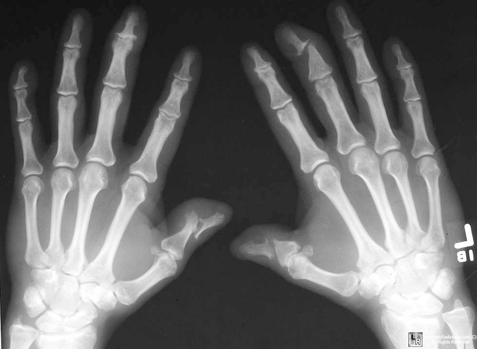
- DIP involvement, including the classic "pencil-in-cup" deformity (as seen above in top image)
- marginal erosions with adjacent bony proliferation ("whiskering")
- small-joint ankylosis (loss of space)
- osteolysis of phalangeal and metacarpal bone, with telescoping of digits (second image above)
- periostitis and proliferative new bone at sites of enthesitis.


3) Physical Examination for Axial Involvement: 
To Assess the sacroiliac joint: 
- palpation along SI joint
- FABERE test: flexion, abduction, external rotation and extension of affected leg (figure 4). Test is positive if tested leg cannot be lower parallel to other leg- this will occur with hip disease, iliopsoas spasm or SI joint disease.
- lateral compression of pelvis
- Gaenslen test (below):


To Assess mobility of lumbar spine:
- Modified Schober test: mark the point 10cm above and 5cm below the level of the dimple of Venus at the midline. Measure again with full forward flexion. This should increase to at least 20cm in flexion.
- standing back to a wall: distance from fingertip to floor on lateral flexion


4) BONUS! Diagnostic Criteria:

CASPAR criteria, a patient must have inflammatory articular disease (joint, spine, or entheseal) with 3 points from any of the following five categories:
  1. Evidence of current psoriasis, a personal history of psoriasis, or a family history of psoriasis
  2. Typical psoriatic nail dystrophy observed on current physical examination
  3. A negative test result for rheumatoid factor
  4. Either current dactylitisf or a history of dactylitis recorded by a rheumatologist
  5. Radiographic evidence of juxtaarticular new bone formation in the hand or foot

    That's all folks!


Wednesday, November 14, 2012

Myasthenia Gravis


Today in morning report we heard about a patient with dysarthria, dysphagia and ptosis. The diagnosis was Myasthenia Gravis.

Pathophysiology: 
- antibodies bind to the post-synaptic acetylcholine receptors
- this blocks acetylcholine binding, lead to crosslinking of receptors promoting internalization and degradation. 
- thus the number and availability of receptors are reduced
- thus end-plate potentials are insufficient to generate action potentials in a number of muscle fibers, causing weakness.

Presentation: 
- Up to 65% of patients initially have ocular symptoms of double vision and drooping of the eyelids.Less than 1/4 present with bulbar symptoms (dysarthria, dysphagia, altered voice). 
- Symptoms are typically better in the morning and after rest. 

Most Useful on history and physical:
- To rule in: “speech becoming unintelligible during prolonged speaking” (LR 4.5), peek sign (LR 30- illustrated in the third frame above), but their absence does not rule it out
- To confirm diagnosis or rule it out:  ice test (positive LR 24, negative LR 0.16), sleep test (positive LR 53, negative LR 0.01) or edrophonium (acetylcholinesterase inhibitor) test (positive LR 15, negative LR 0.11)

Confirming the diagnosis:
1. Acetylcholine receptor antibody test: most specific for MG. Sensitivity in generalized MG is 80%-96%,  but up to 50% of patients with purely ocular myasthenia are seronegative.

2. Single-fiber electromyography: highly sensitive for disorders of the neuromuscular junction but is not specific for myasthenia gravis.

Management:
- Rule out associated condition such as thymoma with CT chest and hyperthyrdoidism with TFTs. 
- Work up for other inflammatory conditions (ANA, RF)
- PFTs to assess for respiratory muscle weakness 

- Symptomatic treatment with pyridostigmine
- Indications for thymectomy: all patients with thymoma, some patients without thymoma (if generalised MG and under age 55 as 85% will have an improvement... however benefit takes years)
- Immunosuppression with glucocorticoids, azathioprine and others
- Avoid exacerbating drugs (including magnesium, beta-blockers, quinolones and macrolides)

Myasthenia Crisis:
- Myasthenic crisis is defined as an exacerbation of weakness sufficient to endanger life
- Close monitoring is key: Vital capacity (VC) and/or maximal inspiratory force (MIF) should be measured frequently, as often as every two hours. May need BiPAP or intubation if VC falling.
- Treat infectious triggers
- Immunomodulator: start prednisone or other immunosuppressive. With steroids, consider slow up-titration as high doses of steroids can exacerbate weakness.
-  IVIG 2g/kg over 5 days- improvement expected within a week
- Can also use Plasmapharesis but this is less well studied than IVIG
- Some advocate holding the anticholinesterase medication as "cholinergic crisis" can contribute to weakness

Tuesday, November 6, 2012

Myocarditis


Today in morning report we heard about a young woman with myocarditis.

Here were some key points:

1) Dangerous causes of chest pain in a young person:
Cardiac: 
- pericarditis
- myocarditis
- myocardial ischemia from drug use, thrombophilia, coronary artery dissection or premature atherosclerosis
- aortic dissection with condition predisposing to aortopathy such as Marfan's, Ehler's-Danlos, Ankylosing spondylitis or bicuspid aortic valve
Lung:
- pulmonary embolism
- pneumothorax
GI:
- Esophageal rupture

2) Causes of Myocarditis:
- Viral and post-viral: mostly Coxsackie, adenovirus, parvovirus and herpes viruses
- Bacterial: Borrelia burgdorferi and Ehrlichia species
- Parasites: Babesia, Trypanosoma cruzi
- Toxic: alcohol, radiation, chemicals (hydrocarbons and arsenic), and drugs, including doxorubicin
- Hypersensitivity: sulphonamides and penicillins
- Idiopathic: giant-cell myocarditis (autoimmune) and cardiac sarcoidosis

3) Complications of Myocarditis
- heart failure and cardiogenic shock
- arrythmias including high degree heart block
- sudden cardiac death as a result of either of the above


Check out this negative trial of cyclosporine and azathioprine for myocarditis.
Here is a great review of Myocarditis

Friday, November 2, 2012

Lower Back Pain


Today in morning report we talked about lower back pain.

Here are some pearls from the discussion:

1) Red flags are reasons to do more advanced imaging (ie. CT or MRI). They are also clues that you may be dealing with something like infection or malignancy. These include:
- age over 60
- night pain ("can't find a comfortable spot in bed")
- fever, other constitutional symptoms
- history of malignancy
- IVDU
- bowel and bladder incontinence

2) Neurological exam for radiculopathy- in Dr Carette's word "it's all in the feet".
A patient with back pain may be limited by pain in terms of what they are willing to do at the hip and knee. The neurological assessment of the foot can be very high yield, with some useful tricks as follows:

- Motor testing: to test L5 only, test extensor hallucis longus with resisted big toe extension. Test S1 with resisted toe curling.

- Sensory: test sensation of medial aspect of shin (L4), in web between big toe and D2 (L5) and on lateral foot and sole (S1)

- Reflexes: Patellar (L2-4) and Ankle jerk (S1).

Note if you want to test L5, can check for the hamstring reflex  as follows: The patient should be lying in the supine position with the knee and hip partially flexed and the leg supported by the examiner's hand. Then, using your reflex hammer, tap one of the medial hamstring tendons behind the knee, causing contraction of the tendon and flexion of the knee.

Follow this link to great review of low back pain from NEJM

Tuesday, September 18, 2012

Pneumocystis jirovecii Pneumonia


Today in morning report we discussed the diagnosis and management of PJP.

Check out this previous blog post on the subject.

Note that workup of a patient with suspected PJP includes an artieral blood gas on room air to determine if criteria are met for treatment with steroids. These are:
- PaO2 less than 70 mmHg
- Aa gradient greater than 35 mmHg

If oxygen saturation is low by pulse oximetry, this would also qualify the patient for steroids.

Friday, September 14, 2012

Vertigo


Today we talked about a patient with vertigo who ended up having something really rare.

It gave us a chance to revisit the often tricky approach to vertigo.

Here are some important points:

1) Key question is distinguishing a peripheral from a central cause of vertigo, as the central causes are the most dangerous. There are some clues on history and physical exam that can help:
History: 
- Stroke risk factors: central
- Presence of other neuro symptoms: central
- Presence of other ear-related symptoms: peripheral

Physical:
- Vertical or gaze-evoked (ie changes direction depending on where patient looks) nystagmus: central
- Nystagmus suppresses with visual fixation: peripheral. Note: would also increase when fixation removed, can test this by using fundoscopy, nystagmus seen with ophthalmoscope to increase when you cover the other eye
- Severe unsteadiness (even with eyes open): central (note in peripheral may tilt to side of lesion, romberg positive)
- Any other neuro findings: central

2) Consider brain imaging even if history and physical suggest a peripheral cause in the following cases:
- older patient
- stroke risk factors
- and acute onset vertigo that persists for more than 48 hours

MRI/MRA are needed in this case since CT is not great for our areas of interest: the brainstem and cerebellum.

 Note that a labyrinthine TIA/stroke will look like a peripheral lesion on history and physical.

3) The image above probably depicts something similar to the Dix-Hallpike maneuver, used to diagnose Benign Paroxysmal Positional Vertigo.

Follow this link to see a video of the nystagmus seen in such patients.

Follow this link for a great NEJM review of acute vertigo. It's an oldie but a goodie.

Wednesday, September 12, 2012

Infective Endocarditis


Today we had a very high yield morning report on infective endocarditis.

Here is a recap of some important points from the discussion:

1) The Modified Duke's criteria is used to make the diagnosis.
Diagnosis is made by any of the following:
- pathology confirmation
- presence of 2 major criteria
- presence of 1 major and 3 minor criteria
- presence of 5 minor criteria

Possible endocarditis: 1 major and 1 minor OR 3 minor criteria are met.

Major Criteria:
A) Micro criteria (any of the following)
a) Culture of a typical endocarditis organism (in 2/2 cultures)
- Staph Aureus
- Viridans group Strep
- Strep gallolyticus (formerly Bovis) and nutritionally-variant strep
- HACEK organism
- Community-acquired enterococci (no primary focus)

b) Single positive culture of Coxiella Burnetti (Q fever) on culture or serology IgG positive >1:800

c) Persistently positive cultures with another organism: 3/3 positive or 3/4 positive.

B) Evidence of cardiac disease: do TTE first, then TEE if prosthetic valve OR at least possible I.E. by clinical criteria. Any of:
- vegetation
- abscess
- new partial dehiscence of prosthetic valve
- new regurgitant murmur

Minor Criteria: 
- Vascular phenomena: splinter hemorrhages, conjunctival hemorrhages, Janeway lesions,
- Embolic phenomena: Glomerulonephritis, RF positivity, Osler's nodes, Roth spots
- fever of at least 38 degrees
- predisposing condition: IVDU, congenital cardiac disease or prosthetic valve
- positive cultures not meeting descriptions above.

2) When is prophylaxis for endocarditis needed? In a high-risk patient AND a high-risk procedure
High risk patients:
- past IE
- prosthetic valve
- uncorrected cyanotic shunts, or in first 6 months post correction
- heart transplant valvulopathy

High risk procedure:
- dental with gingival manipulation
- transbronchial biopsy
- involving infected GI, GU, skin, muscle

3) When to consult cardiac surgery in endocarditis:
- refractory CHF or shock
- uncontrolled infection: abscess/fistula, ongoing fevers, ongoing culture positivity, or fungal or resistant organism
- very large vegetation (>1cm)
- reucrrent embolic phenomena


Check out this very recent small study published in NEJM which sheds some light on those "debatable" indications for cardiac surgery (ie. size of vegetation and embolic phenomena). In this study, patients randomised to early surgery had significantly fewer embolic events (most of these were strokes) than those who had conventional therapy. Of note, 77% of patients on the conventional therapy group went on to have surgery (just later) and there was no difference in 6 month mortality in the two groups.

Here is a link to the classic NEJM review on endocarditis 


Thursday, September 6, 2012

Hepatorenal Syndrome


Today in morning report we heard about a patient with decompensated liver disease. He presented with jaundice and an elevated creatinine.

When patients with cirrhosis present with an AKI, we ask is this Hepatorenal syndrome or something else?

Things that can trigger renal failure in cirrhotic patients:
- Overdiuresis
- NSAIDs (cirrhotic patients very dependent on renal prostaglandin synthesis) and other nephrotoxins
- GI bleed
- Bacterial infections including translocation of bacteria in gut and spontaneous bacterial peritonitis (SBP)
- Any acute decompensation of cirrhosis

Hepatorenal Syndrome is "characterized by functional renal vasoconstriction that leads to a severe reduction in GFR with minimal renal histologic abnormalities" (NEJM, see below). The figure above shows which mechanisms lead to this.

Type 1: Develops rapidly. Creatinine doubling over less than 2 week period
Type 2: More gradual than type 1.

To be labelled hepatorenal syndrome:
- no improvement after 2 days off diuretics and albumin 1gm/kg/day for 2days minimum
- no recent nephrotoxic drugs
- no shock
- no evidence of renal parenchymal disease (significant proteinuria, hematuria, or abnormal looking kidneys on ultrasound)

Initial Management of AKI in patient with ascites:
- Exclude bacterial infection: diagnostic paracentesis + other cultures
- Exclude GI bleed
- Hold diuretics
- Stop nephrotoxic drugs
- give a trial of albumin as above

Distinguishing hepatorenal syndrome from ATN can be tricky as both can cause the presence of heme granular casts in the urine. Once diuretics are held, a FENa (fractional excretion of sodium) less than 1% supports the diagnosis of hepatorenal syndrome.

Specific therapies for hepatorenal syndrome:
- Terlipressin: improves renal function, no effect on survival found so far
- Midodrine and octreotide together: small studies suggest benefit on renal function
- TIPS= transjugular intrahepatic portosystemic shunt: some observational studies show improvement, however many patients with HRS too sick to undergo this procedure
- Liver transplantation- don't forget to consider this

Prognosis of HRS is very poor. Mortality is higher with type 1 hepatorenal syndrome than with type 2 (median survival, 1 month vs. 6 months)

Prevention of HRS in patients with ascites:
- Albumin as part of treatment for SBP: 1.5 gm/kg at initiation and 1gm/kg at 48 hours.
- Prevention of SBP with norfloxacin in selected patients- see this study


Here is a link to the most recent NEJM review on the topic

Tuesday, September 4, 2012

Flash Pulmonary Edema


Today in morning report we touched on the causes of flash pulmonary edema. We haven't addressed this on the blog previously, so I thought we would go over it:

Flash pulmonary edema is a particularly sudden form of Acute Decompensated CHF (in patient without chronic CHF). Mechanism of flash pulmonary edema is a sudden increased in LV end-diastolic pressure

Classic causes of flash pulmonary Edema:

1) Primary Cardiac Problems
- Mitral stenosis: in setting of tachycardia, atrial fibrillation, or high circulating volume of pregnancy
- Acute mitral or aortic regurgitation
- Arrythmia in a patient with pre-existing diastolic dysfunction
- Sudden Pump failure: acute MI or stress-induced cardiomyopathy

2) Hypertensive Emergency

3) Renovascular
Bilateral renal artery stenosis more commonly than unilateral. Patient with RAS are predisposed to developping flash pulmonary edema because of "(i) defective natriuresis; (ii) increased haemodynamic burden and exacerbation of diastolic dysfunction and (iii) failure of the pulmonary capillary blood–gas barrier" (Messerli FH. Eur Heart J32 (18): 2231-2235). 


Acute Treatment is centered around decreasing preload as well as afterload:
- Lasix
- (Morphine) Limiting supporting data. In theory, decreased sympathetic drive, thus leads to veno/arterial vasodilatation
- Nitroglycerin (venodilator thus reduces preload,  weaker arterial vasodilator)
- Oxygen
- P= Position (sitting up) but also Pressure (ie. Non-Invasive Positive Pressure Ventillation aka CPAP or BiPAP)

Have a great day!

Thursday, August 30, 2012

Heart Failure with Preserved Ejection Fraction

 Today's morning report was about a man who developed flash pulmonary edema in the context of an NSTEMI.

Follow this link for a great CMAJ review of the management of acute decompensated heart failure. 


One thing that was touched on was the concept of congestive heart failure with preserved ejection fraction, aka "Diastolic Dysfunction" which occurs in 1/3 of patients with heart failure.

This type of dysfunction occurs as a result of concentric LV hypertrophy, and impaired ventricular relaxation as a result. As the ventricle does not sufficiently relax in diastole, filling (and thus forward flow) are impaired.

Risk factors include age, female gender, hypertension, coronary artery disease, diabetes mellitus, restrictive cardiomyopathy from infiltrative causes.

Diagnosis: Clinical evidence of heart failure with an ejection fraction greater than 50%. Echocardiogram can be helpful as it may show alterations in the E:A ventricular filling ratio (E is early, A is for atrial kick), as well as left atrial enlargement (more than 4cm), or evidence of hypertrophy (IV septum greater than 1cm, LV mass greater than 100gm/m2 body surface area).

Treatment: There is a lack of evidence when it comes to treating CHF with preserved EF. Guidelines (ACC/AHA) recommend controlling blood pressure, treating edema and controlling heart rate to allow greater LV filling time.


Follow this link for a NEJM review on the subject. 

Cellulitis and mimickers


Yesterday's Morning Report was on cellulitis and mimickers.

One key point made was the distinction between cellulitis and erysipelas, which is an infection of the superficial skin layers caused by beta-hemolytic streptococci. Unlike cellulitis, the borders of erysipeloid areas are very well demarcated. 

From a previous morning report on cellulitis:

Predisposing factors: saphenous vein harvesting, venous insufficiency, mastectomy with lymph node dissection, liposuction, "skin popping" in IVDU

Source: portal of entry in skin (e.g. tinea pedis, ulcer)- by far most common; other possibilities include osteomyelitis, bacteremia. 
Unusual sources: seawater (vibrio vulnificus), fresh water (aeromonas hydrophilia), fish (strep iniae)

Micro: 
80% gram +ve (staph, strep), 20% gram -ve. Aspirates/swabs are not indicated (unless ulcer) 
Broader coverage may be indicated in pts with DM2. 
Blood cultures are indicated in lymphedema, buccal, periorbital, water exposure, chills or fever. Otherwise, bacteremia is rare (less than 4%).

Empiric treatment: Cefazolin or cephalexin. Other possibilities: cloxacillin, clindamycin, penicillin, amoxicillin-clavulin. May want broader coverage (e.g. gram -ve coverage) in diabetics

Ancillary measures: Elevation, immobilization. Interdigital dermatophytic infections should be treated (e.g. clotrimazole and miconazole), terbinafine, etc
Click here for a review of cellulitis from NEJM

Important Conditions that Masquerade as Cellulitis:

Infectious:
- Necrotising fasciitis/ Clostridial myonecrosis

Non-infectious:
- Just plain edema with or without changes of chronic venous stasis- highly probable if bilateral
- Lymphedema
- Lipodermatosclerosis (in patients with chronic venous stasis)  fibrosing panniculitis characterized by advanced hyperpigmentation and induration involving most of the leg circumferentially.
- Superficial thrombophlebitis/DVT
- Contact dermatitis
- Systemic drug reaction
- Gout: in addition to arthritis, often have overlying skin induration and involvement of tendons
- Sweet's syndrome: acute febrile neutrophilic dermatosis associated with AML
- Well's syndrome aka eosinophilic cellulitis: urticarial lesions, transient systemic eosinphilia.


Click here for a great Annals review of diseases that masquerade as cellulitis. 

Wednesday, August 22, 2012

Staphylococcus Aureus and other goodies


Today we talked about a woman with a paraspinal abscess and Staphylococcus Aureus in the blood.

Some pearls of the discussion were:

- Causes of paraspinal/epidural abscess include: IVDU, endocarditis, contiguous spread from GI/GU infections

- Psoriasis is a risk factor for SA bacteremia. These patients should also have their psoriasis treated (note psoriatic skin lesions do not count as a removable source of infection)

- Psoas abscess is often a clue that there is a vertebral infection (as the infection spreads into the psoas from the vertebral/paravertebral space). Thus, if patient has a psoas abscess, consider getting a spine MRI as well.

- Staph Aureus bacteremia should be treated with 4-6 weeks of IV antibiotics unless all of the following conditions are met in which case can treat for 2 weeks (based on this 2003 Archives paper):

1. removable source
2. no evidence of metastatic infection
3. Resolution of fever by 72 hours of therapy
4. Negative blood cultures by 48-96 hours

For more on Staph Aureus bacteremia see this previous blogpost

For more on epidural abscess, see this previous blogpost as well as this NEJM paper 


Thursday, August 9, 2012

Vasculitis


Dr Simon Carette took us through the approach to vasculitis as we discussed a patient with a case of Microscopic Polyangiitis (MPA).

This patient presented with mononeuritis multiplex as well as purpura of the legs.

Some pearls that he shared:

1) Mononeuritis multiplex: dysfunction of multiple named peripheral nerves. Pathognomonic for vasculitis. The most common causes are:
- Polyarteritis Nodosa
- Churg-Strauss Syndrome
- Cryoglobulinemia
- Connective Tissue Disease-Associated (note that not all CTDs cause vasculitis- the ones that do most commonly are lupus, rheumatoid arthritis and Sjogren's).

Mononeuritis mulitplex could, for example, present as an isolated foot drop. One important question when examining these patients is: how to distinguish a common peroneal nerve palsy  from an L5 radiculopathy (both cause foot drop).
Key distinction: Ankle inversion and thigh abduction are weak in L5 radiculopathy, but not in common peroneal nerve palsy. Weak dorsiflexion and eversion are seen in both conditions.

2) Skin lesions in vasculitis: presentation depends on which vessels are involved.
- Small vessel vasculitis- superficial vessels involved= petechiae, purpura (see lower image above), hemorrhagic bullae.
- Medium-sized vessel vasculitis- depper vessels involved= livedo reticularis (classic in PAN, see top image above), nodules, ulcers. Note that a standard punch biopsy will not reach the deeper layers and miss the diagnosis. In the presence of these skin lesions, a deeper punch or wedge biopsy is needed.

3) ANCA Antibody Testing:
- c-ANCA highly specific for anti-PR3 antibody and Granulomatosis and Polyangiitis (previously known as Wegener's)
- p-ANCA much less specific for anti-MPO- thus positivity on p-ANCA testing should prompt a follow-up ELISA test for anti-MPO (many other things including IBD can give you a positive p-ANCA that are not associated with vasculitis). If anti-MPO positive, 80% specific for a vasculitis (MPA, Churg-Strauss)


Finally, check out this NEJM CPC case on PAN for a good review of the approach to a patient with vasculitis.

Tuesday, August 7, 2012

Tamponade or PE?


Today we discussed a tricky case of a patient with both a pericardial effusion and a pulmonary emobolism. When they developed shock, the question became is this tamponade or a massive PE?

On physical exam, both groups of patients will have:
- sinus tachycardia
- hypotension
- high JVP

In tamponade: muffled heart sounds, pericardial friction rub, pulsus paradoxus

In massive PE: signs of DVT

Note that an increased pulsus (ie>10 mmHg) can also be seen in other conditions: profound hemorrhagic shock, obstructive lung disease... and massive pulmonary embolism!

In an unstable patient, bedside investigations would include an ECG...

ECG findings of tamponade: 
- sinus tachycardia
- low voltages (ie less than 5 in limb leads, less than 10 in precordial leads)
- pericarditis findings: ST elevation, PR depression
- electrical alternans (beat-to-beat variation in QRS amplitude, caused by swinging of the heart in the pericardial fluid) - rare but very specific.

ECG findings of pulmonary embolism:
- sinus tachycardia
- atrial arrhythmias
- S1Q3T3-rare but specific ECG pattern
- iRBBB/RBBB
- RAD
- non-specific ST/T wave changes
- precordial T-wave inversion (Rt heart strain pattern)

To definitively sort these two out, an echocardiogram is needed. 

Echocardiogram findings of massive PE:
- Increased RV size
- Decreased RV function
-Tricuspid regurgitation

Echocardiogram findings of tamponade:
- RA/RV diastolic collapse
- ventricular interdependence: reciprocal respiratory variation in volume in right and left heart, as well as flows across AV valves (as in figure above).
- IVC full, collapses by less than 50% on inspiration

Check out this NEJM review on Acute Cardiac Tamponade

Friday, August 3, 2012

Kidney injury and ACE inhibitors

Today in morning report we touched on the perils of volume depletion in patients who are taking ACE inhibitors. Have you ever wondered why this situation would precipitate acute kidney injury?


The issue is this: ACE inhibitors offload the glomerulus (and thus protect it from wear-and-tear) by causing efferent arteriolar dilatation. Normally in situations of volume depletion, the Renin-Angiotensin system is activated, and angiotensin II causes efferent arteriolar constriction, thus increasing the pressure in the glomerulus (think of it as a "squeeze") and thus preserving GFR. If you have a ACE inhibitor (or ARB) on board then you don't get the efferent constriction, thus no squeeze, thus drop in GFR. This causes AKI.

Patients on ACE inhibitors should be warned about this complex issue. While the ACE inhibitor protects their kidneys in the longterm, it can hurt them in the short term if your patients takes his ramipril while volume depleted. So please hold it if there is diarrhea, vomitting, decreased PO intake, bleeding etc.

Cheers! Have a great long weekend!

Wednesday, August 1, 2012

Secondary Causes of Hypertension & Hypertensive Emergencies


When to consider a secondary cause of hypertension:
1. sudden onset or worsening of hypertension at any age
2. onset of hypertension in those less than 30 years old (with no family history or obesity)
3. hypertension resistant to 3 drugs


Secondary causes of hypertension and their clues:
-Renovascular disease: abdominal bruit, rise in creat>30% upon ACE inhibitor or ARB initiation, atherosclerosis elsewhere, history of flash pulmonary edema with hypertensive episodes

-Pheochromocytoma: paroxysmal hypertension, typical spells (headache, palpitations, sweating, panic attacks, pallor), hypertension triggered by beta-blockers, MAO inhibitors or changes in abdominal pressure (ie. intraoperatively),adrenal mass

-Hyperaldosteronism (often missed!): hypokalemia  less than 3 .5 without diuretics or less than 3.0 on diuretics, adrenal incidentaloma on imaging

-Cushing's syndrome: typical appearance, history of exogenous steroids

-Sleep apnea: body habitus (including neck circumference more than 16 inches in women and more than 17 inches men), history of snoring/apneic spells/morning headache/daytime somnolence

-Medications: OCP, HRT, some NSAIDS, some antidepressants (eg Venlafaxine), sympathomimetics including decongestants

-Coarctation of the aorta: don't forget to check for brachiofemoral delay in this patients... has been detected for the first time in adulthood

-Hypothyroidism

-Hyperparathyroidism


Hypertensive Emergencies
"Urgency": SBP over ~180 or DBP over ~110 without end-organ damage- needs correction over days with oral agents
"Emergency": Above, but with acute end-organ damage, needing urgent lowering, usually with IV medications in a monitored setting.

End organ complications and specific treatments:
1) Aorta- dissection (B-blockade, nitroprusside after B-bl. No pure vasodilators)
2) Brain- encephalopathy (note that headache without neuro deficits does not count!)- sz, cerebral hemorrhage/infarction, raised ICP
3) Heart- MI, CHF (acute diastolic dysfunction leading to pulmonary edema)- careful with B-bl. May use nitro infusion
4) Kidney- renal failure- careful diuresis, calcium antagonists useful
5) Placenta (pre-eclampsia)- hydralazine, labetalol, delivery
6) Hemolysis (can look just like TTP with MAHA, fragments)
7) Eyes: papilledema, hemorrhages

Treatment Targets: 
-         emergency: lower BP by no more than 25% in minutes to 2 h using IV medications (exception: aortic dissection, where it must be lowered more rapidly)
-         urgency: lower BP over hours to days with PO
-         meds PO: amlodipine, captopril, hydralazine, clonidine
-         meds IV: labetalol, nitroprusside, nitroglycerin, hydralazine


Tuesday, July 31, 2012

Sickle Cell Disease


Today we heard about a patient with a sickle cell crisis.

Much has been written on the topic on this blog.

Check out a blogpost on Sickle cell anemia here

Acute chest syndrome is a dreaded complication, read all about it here

Monday, July 30, 2012

Pyogenic Liver Abscess


Today we discussed a case of pyogenic liver abscess. This is a very hot topic right now! Incidence increases with age, which may explain why we are seeing so many of these on general internal medicine wards. They most commonly involve the right lobe of the liver, as it is larger and receives more blood supply.

Causes of pyogenic liver abscess can be categorized as follows:

1. Biliary tract disease: ascending cholangitis caused by obstruction (stone, pancreatic or cholangioCA, stricture)

2. Portal vein seeding as a result of intraabdominal disease: this can be secondary to appendicitis, diverticulitis, bowel perforation, malignancy. May be accompanied by pyelephlebitis (suppurative thrombophlebitis of the portal vein).

3. Hepatic artery seeding: from systemic bacteremia with or without bacterial endocarditis

4. Contiguous focus of infection: gallbladder disease, subphrenic or perinephric abscess.

5. Traumatic: liver laceration, post-resection, post radiofrequency ablation, migrated foreign body (eg: fishbones through gastric antrum).

5. "Primary"=cryptogenic: Hypermucoviscous variants of Klebsiella pneumoniae are increasingly being recognized as a cause. This is seen most commonly in patients of Southeast Asian descent and has been associated with diabetes mellitus.

Microbiology of liver abscesses: Many are polymicrobial (especially if secondary to biliary tract disease or intraabdominal infections). Common pathogens are: E.Coli, Klebsiella pneumoniae, Strep (including Strep anginosus group- famous for abscesses), anaerobes.

Treatment is focused on source control=drainage of abscess. Broad-spectrum antibiotics (eg:ceftriaxone,flagyl) are used until a specific pathogen is isolated from drainage or blood cultures (positive 50% of the time). Antibiotics are initially intravenous, then stepped down to oral for a total 4-6 week course.

Check out this recent case from TGH with a review on the topic by our very own Wayne Gold and Derek MacFadden: Here

Friday, July 27, 2012

DKA Management


Above is a useful algorithm for the management of DKA, taken from CMAJ's 2003 paper (a classic and must-read for medicine residents):

Diagnosis and treatment of diabetic ketoacidosis and the hyperglycemic hyperosmolar state 


Added management pearls:

- Many people find it convenient to manage DKA by a flowchart updated ~hourly (although not everything needs to be checked hourly). You may want to record:
Time, Vitals, urine output, pH, HCO3, AG, Ket, Glu, K, PO4, IV fluid rate, insulin dose


-Ketones may increase even though the anion gap is decreasing. This is because beta-hydroxybutyrate (major ketoacid of DKA) is not detected by the ketone test. It is then metabolised to acetoacetate, which is detected.


-When calculating the anion gap, use the uncorrected sodium concentration. Note that even though we see hyponatremia in hyperglycemic states (and there is a "correcting calculation"), the anion gap reflects the balance between positively and negatively charged electrolytes in the extracellular fluid. Glucose is electrically neutral and does not directly alter the anion gap. However, glucose is osmotically active so water is pulled into the extracellular fluid. This has a dilutional effect on all extracellular electrolyte concentrations, both positive or negative, and so the anion gap is minimally altered.

- Use of bicarbonate is reserved for severe acidosis (pH<7.0) after 1 hour of rehydration

- Expect to see non-anion gap metabolic acidosis (ie. hyperchloremic) after AGMA resolves, because of loss of ketone bodies without H+ (i.e. loss of anion with Na+/K+ as cation). This is equivalent to the loss of a "potential bicarbonate". This lost bicarbonate is replaced by chloride ion (which is abundant in the saline we are giving).

Thursday, July 26, 2012

CN IV Palsy


Check out this Right CN IV Palsy- note that because CN IV innervates the superior oblique, the affected eye cannot look down and in - the patient will report improvement of diplopia with a corrective head position: tilting the head toward the opposite shoulder 

Tuesday, July 24, 2012

PFO/Stroke in the Young


This morning we discussed a case of stroke in the young. See a previous blog post on stroke in the young here.

Two specific points with respect to this came up:

1) In patients with cryptogenic stroke, does PFO closure prevent recurrent strokes?

Answer: in a recent (NEJM 2012) unblinded multicenter study of 909 patients (ages 18-60yrs) randomised to closure or medical management (antiplatelet or anticoagulation at physician's discretion), there was no significant difference in the primary endpoint (composite of stroke or TIA in 2 years, death of any cause in 30 days, and death from neurological cause at 31 days to 2 years). See the study here.


2) One question that this brings up for me: does anticoagulation prevent recurrent strokes in patients with PFOs?

Answer: data and expert opinion are conflicted as to the optimal medical management of a patent foramen ovale in a patient with an otherwise cryptogenic stroke. In this study subgroup, patients were randomised to ASA 325mg or warfarin following stroke (250 cryptogenic out of a total 601 strokes). Of note, PFO was found in 39.2% (98/250) of patients with cryptogenic stroke, compared with 29.9% (105/351) in patients with known cause of stroke. Note that this is not a huge difference in prevalence between the two groups.

With respect to treatment, there was no significant difference in the time to recurrent ischemic stroke or death between those treated with warfarin and those treated with aspirin (P=0.49; hazard ratio 1.29; 95% CI 0.63 to 2.64; 2-year event rates 16.5% versus 13.2%).

There is some data suggesting that the presence of an atrial septal aneurysm along with a PFO confers greater risk.

The AHA Guidelines from 2011 are as follows:

- For patients with an ischemic stroke or TIA and a PFO, antiplatelet therapy is reasonable (Class IIa; Level of Evidence B). 

- There are insufficient data to establish whether anticoagulation is equivalent or superior to aspirin for secondary stroke prevention in patients with PFO (Class IIb; Level of Evidence B). (New recommendation)
 
- There are insufficient data to make a recommendation regarding PFO closure in patients with stroke and PFO (Class IIb; Level of Evidence C)

3) Finally: What the heck is CADASIL?

Answer from Harrison's:  
-  cerebral autosomal dominant arteriopathy with subcortical infarcts and leukoencephalopathy (CADASIL)
- inherited disorder that presents as small-vessel strokes, progressive dementia, and extensive symmetric white matter changes visualized by MRI.  
- 40% of these patients have a history of migraine with aura. 


See you tomorrow!


Monday, July 23, 2012

A tale of 2 cases - July 23, 2012


Thank you to team 8 for bringing 2 cases, and to Dr. Wayne Gold for hosting today’s morning report.  We discussed 2 interesting cases.

The first case involved a patient with a stroke.  We discussed that in patients already receiving ASA, neither Plavix (clopidogrel) nor Aggrenox (ASA + extended release dipyridamole) is superior to the other agent for preventing recurrent stroke, as demonstrated in the PRoFESS trial (Prevention Regimen for Effectively Avoiding Secondary Strokes) published in 2008 (Sacco RL, et al; PRoFESS Study Group. Aspirin and extended-release dipyridamole versus clopidogrel forrecurrent stroke.  N Engl J Med. 2008 Sep18;359(12):1238-51.).  They randomized 20322 patients and followed them for a mean of 2.5 years for the primary outcome of first recurrence of ischemic stroke.  No difference was found in the primary outcome (9% in the ASA + extended release dipyridamole group and 8.8% in the clopidogrel group; hazard ratio 1.01; 95% CI 0.92 to 1.11).  However, some patients may benefit from anticoagulation if there is known cardioembolic stroke risk such as atrial fibrillation, or apical thrombus.  In reality, the benefit of antiplatelet or anticoagulant therapy must be balanced against the individual bleeding risk of the patient.

We then discussed the differential diagnosis of episodic decreased level of consciousness post stroke.  The most prominent being post-stroke seizure (+/- accompanying post-ictal phenomenon).  Hemorrhagic conversions are less likely to present as transient decreased level of consciousness.  It is important to obtain imaging to rule out structural causes, EEG to assess for seizure, and to complete a metabolic work-up to rule out non-CNS causes.  Syncope is also in the differential diagnosis of episodic loss of consciousness and cardiac investigations (including the ruling out of dysrhythmias) may be warranted.

In the discussion of our second case, we discussed the principles of management of febrile neutropenia.  We discussed the importance of starting empiric broad-spectrum antibiotics first without waiting for results of cultures.  Minimum of blood and urine cultures, and chest x-ray should be obtained.  Other investigations should be directed based on patient’s presenting symptoms and physical exam findings.  Empiric therapy should cover gram positive organisms (including Staph aureus) +/- CNST (coagulase negative staph), AND gram negative organisms (including Pseudomonas).  Many institutions have empiric therapy guidelines based on local resistance pattern.  The Infectious Diseases Society of America (IDSA) Guideline on antibiotics used in febrile neutropenic patients can be found here.  (Freifeld AG. Clinical practice guideline for the use of antimicrobial agents inneutropenic patients with cancer: 2010 Update by the Infectious DiseasesSociety of America.  Clin Infect Dis. 2011 Feb 15;52(4):427-31.)

We then discussed a rash in the setting of febrile neutropenia.  In general, the rash can represent a primary skin/soft tissue infection, or a secondary manifestation of a systemic infection.  The latter can be from hematogenous seeding (as in the case of Janeway lesions in infective endocarditis), immune mediated (e.g. disseminated gonococcal infection), activation of coagulation cascade (purpura fulminans), or toxin-mediated (as in staphylococcal toxic shock syndrome).  In our patient, we suspect the skin lesion is ecthyma gangrenosum.  In febrile neutropenic patients, this may represent a gram negative infection (commonly Pseudomonas).  Other infections (e.g. fungi [Candida, Aspergillus]) are also possible.  See this article by Yas Moayedi which summarizes mechanism of skin manifestation of systemic infection, other organisms that can cause ecthyma gangrenosum, and principles of management.  (Moayedi Y,Bunce PE, Sade S, Ghazarian D, Gold WL.  Fromthe outside looking in.  Am J Med. 2012May;125(5):457-60.).

Note that in most patients with skin infection, we do not routinely obtain aspirate and biopsy of the skin.  However, in a patient with febrile neutropenia, this is worthwhile as it has a higher yield of recovering the organism(s) responsible.  We chose to treat this patient with two anti-pseudomonal agents to ensure that there is at least one agent that is active against the organism should it later turn out that this patient had a resistant organism.  Once sensitivity known, antibiotics can then be tailored.


Your chief resident (Lauren) will return tomorrow.  Thank you for a week of interesting cases.  Terence

Friday, July 20, 2012

Vasculitis - July 20, 2012


Thank you to team 6 for bringing an interesting case and to Dr. Ho Ping Kong (“As brilliant as House, but nicer.”) for hosting.

Among other things (malignant hyperthermia can be diagnosed with muscle biopsy, nasopharyngeal cancer is EBV associated, etc …), we discussed a case of a woman with likely non-acute kidney disease, rash, and p-ANCA positivity.  This is suspicious for a vasculitic process.  We talked about that imaging (in addition to blood rheumatological markers) is an important part of a vasculitic work-up.  Of course, biopsy can be very helpful as well.

A commonly used classification scheme of systemic vasculitis is the “Chapel Hill” classification, named after the Internal Consensus Conference in 1994 where it was convened for this matter.  It is published in:  Jennette JC, Falk RJ,Andrassy K, Bacon PA, Churg J, Gross WL, Hagen EC, Hoffman GS, Hunder GG,Kallenberg CG, et al. Nomenclature of systemic vasculitides. Proposal of aninternational consensus conference. Arthritis Rheum. 1994 Feb;37(2):187-92.

We will briefly summarize the classification here, but you can get the full details from the actual article:
- Large vessel:  Giant cell (temporal) arteritis, takayasu arteritis
- Medium vessel:  Kawasaki disease, Polyarteritis nodosa
- Small vessel:  Churg-Straus syndrome, Wegener’s granulomatosis, microscopic polyangiitis, cutaneous leukocytoclastic angiitis, essential cryoglobulinemic vasculitis, Henoch-Schonlein purpura.

Churg-Straus, Wegener’s, and microscopic polyangiitis have strong associations with ANCA.

Have a fabulous Friday!