Friday, September 17, 2010

Multiple Myeloma


Today we discussed a classic case of Multiple Myeloma with hypercalcemia, renal failure, anemia, and bone pain secondary to lytic bone lesions... these features make up the mnemonic CRAB: hyperCalcemia, Renal failure, Anemia, Bone pain.

Here is a link to a previous Horses & Zebras blog summarizing the Clinical Features of Multiple Myeloma, as well as some links to articles reviewing investigation and management of hypercalcemia and current management options for multiple myeloma.

Management of hypercalcemia +/- acute renal failure is often the primary problem that needs to be managed on admission to hospital. Hypercalcemia can be considered to be mild (2.6 - 2.9 mmol/L), moderate (3.0 - 3.4 mmol/L), and severe (greater than 3.5 mmol/L); however, there are no official cut-off points for hypercalcemia. As we discussed this morning, ~40% of calcium is bound to albumin and thus low albumin states will lower the measured calcium. This can be corrected roughly by adding 0.2 mmol/L to a calcium measurement for every drop in albumin of ~10.

Treatment of Hypercalcemia

1. First line treatment for hypercalcemia is fluid resuscitation, as the majority of patients will be volume contracted. Rapid, aggressive resuscitation is the goal, monitoring for any potential volume overload/heart failure in susceptible patients.

2. Administration of an IV bisphosphonate is also considered appropriate in severe hypercalcemia. Pamidronate (60 - 90 mg) is the usual choice, given once over 2-4 hours, and most often normalizing calcium within 5-7 days.

3. For more immediate calcium-lowering calcitonin can be considered; however, it is not uncommon for patients to develop tachyphylaxis and thus its use is limited.

4. Alternatively, and often more effective in the setting of lymphoma or multiple myeloma induced hypercalcemia, glucocorticoids such as prednisone (1 mg/kg daily) can be considered.

5. Management of hypercalcemia always includes investigation into the underlying cause, and treatment of such problem otherwise the hypercalcemia will recur.

Friday, September 10, 2010

Guillain-Barre Syndrome

This week we explored 2 different (potential) cases of Guillain-Barre Syndrome, or GBS. First during Physical Examination Rounds and then again in Morning Report.

The most common form of GBS is an idiopathic acute inflammatory demyelinating polyneuropathy (AIDP), and in fact GBS is often synonymous for AIDP. It is an autoimmune process directed against Schwann cell membranes. There are 5 other variants of GBS, including the Miller Fisher Variant which often presents with the triad of ophthalmoplegia, ataxia, and areflexia.

GBS occurs anywhere from 1 - 2 per 100,000 per year. It is often (~2/3 of cases) preceded by either a respiratory or GI infection, most commonly Campylobacter. GBS has also been reported after vaccinations and to be associated with systemic illnesses, such as SLE, Hodgkin's Lymphoma, Sarcoidosis, etc.

Patients with GBS often present with parasthesias in their limbs, followed soon after by ascending weakness. This progresses over days to weeks. GBS can also be associated with autonomic dsyfunction such as orthostasis, urinary retention, constipation, and tachycardia.

Physical examination most often reveals symmetrical motor weakness in an ascending pattern, with absent reflexes, flaccid tone, and a normal sensory exam.

Patients with GBS need to be monitored closely for respiratory failure due to involvement of the diaphragm. A good rule of thumb for consideration of intubation is the 20/30/40 rule:
  • 20: Vital Capacity less than 20 cc/kg
  • 30: Maximum Inspiratory Pressure less than 30 cm H2O
  • 40: Maximum Expiratory Pressure less than 40 cm H2O

Treatment for GBS is most often supportive, and may require an ICU setting for ventilatory support, hemodynamic support, nutritional support, and pain control. If diagnosed within the first 4 weeks then IVIG or plasmapheresis can be considered. A recent Cochrane review suggests that IVIG treatment has similar outcomes to plasmapheresis in the setting of early (ie: within 2 weeks) diagnosed GBS.

Outcomes for patients with GBS are quite good with ~80% achieving full recovery within a few months to a year; however, minor deficits often persist. 5-10% will have permament disabling deficits, and another 2-3% will die as a result of their GBS. Approximately 5-10% of patients may develop one or more relapses of their GBS, leading to a diagnosis of Chronic Inflammatory Demyelinating Polyneuropathy (CIPD).

Here is a very thorough review of GBS from 2005, in The Lancet.

Friday, September 3, 2010

Pulmonary Hypertension


Today we discussed a case of newly diagnosed Pulmonary Hypertension in a young woman.

Pulmonary Hypertension (PH) is defined as a Pulmonary Arterial Pressure over 25 mmHg at rest (normal 12-16 mmHg).

Patients often present with months or years of gradually progressive symptoms, such as shortness of breath, fatigue, cough, angina, peripheral edema, and rarely hemoptysis.

Cardiovascular examination often reveals a loud, palpable P2, a split S2, and an RV heave.

There are multiple causes of PH, but all result in increased pressures within the pulmonary vasculature, either due to vasoconstriction of the blood vessels, obstruction by way of a mass or pulmonary emboli, or fibrosis within the lung parenchyma related to systemic illnesses. In order to overcome this increased pulmonary pressure, the heart then is required to create elevated right-sided pressures in the heart. In turn, this leads to RV hypertrophy and potentially RV failure.

PH is classified according to the WHO into 5 major categories:
Class I: Pulmonary Arterial Hypertension
  • Idiopathic
  • Familial
  • Connective Tissue Disorders, HIV, Drugs, Toxins, Congenital Left-to-Right Shunts, etc.
    Class II: PH with Left Heart Disease
  • Left Atrial or Ventricular Heart Disease
  • Left-sided Valvular Heart Disease
    Class III: PH Associated with Lung Diseases or Chronic Hypoxia
  • COPD, Interstitial Lung Disease, Sleep Disordered Breathing, etc.
    Class IV: PH Due to Chronic Thrombotic and/or Embolic Disease
  • Thromboembolic obstruction of the proximal or distal pulmonary arteries
  • Nonthrombotic obstruction of the pulmonary arteries (ie: tumor, foreign body, etc.)
    Class V: Miscellaneous causes
  • Sarcoidosis, Lymphangiomatosis, etc.

  • Given the numerous potential causes for PH, any patient with suspected PH requires significant investigations to diagnose PH and then to determine the etiology of their PH. This includes basic blood work (looking for evidence of polycythemia), an ECG (evidence of RAE or RVH), a CXR, and echocardiogram, a CT Chest, a V/Q scan (to detect chronic thromboembolic disease), a 6-minute walk test, and a Right-Heart Catheterization to name a few!

    These patients are managed by the multidisciplinary PH team where the goals of treatment include symptom management, improving quality of life, and prolonging survival, which often requires a lung or heart-lung transplant.

    Here is a review article on the mechanisms of PH, and here are the 2009 American College of Cardiology Consensus Guidelines for PH.

    Thursday, July 29, 2010

    TIA Management


    Today we reviewed the approach to a patient presenting with symptoms suggestive of a Transient Ischemic Attack (TIA).

    A TIA is defined as an abrupt focal loss of neurologic function caused by reduction in blood flow that persists less than 24 hours and clears without residual disability.

    Diagnosis is made with a thorough history and physical examination, where the latter can be normal in many patients.

    The patient's risk for progression to stroke can be assessed by the ABCD score, which helps to guide the clinician on the need for admission:

    A: Age - 1-pt if over 60 years old; 0-pt if less than 60
    B: Blood Pressure - 1-pt if SBP greater than 140 OR 1-pt if DBP greater than 90
    C: Clinical symptoms - 2-pts if unilateral weakness; 1-pt if language disturbance but no weakness; 0-pt for other symptoms
    D: Diabetes - 1-pt if patient has diabetes; 0-pt if not
    D: Duration - 2-pts if more than 60 min; 1-pt if 10-59 min; 0-pt if less than 10 min

    Score 0-3: low risk, 2-day stroke risk 1%
    Score 4-5: moderate risk, 2-day stroke risk 4%
    Score 6-7: high risk, 2-day stroke risk 8%

    Low risk patients don't need to be admitted for stroke work-up; however, they do require expedited investigations which sometimes warrant an admission. Moderate risk patients and High risk patients generally warrant an admission and investigations and monitoring for stroke.

    Current Canadian Stroke Guidelines recommend rapid turn around times for investigations in a patient presenting with a TIA. Patients are categorized into Emergent, Urgent, and Semi-urgent; and based on these 3 categories, their stroke work-up is recommended to be performed within the given timeline.

    Emergent: investigations within 24-hrs
    • Symptoms within the previous 24-hrs with 2 or more high-risk clinical features (ABCD criteria)
    • Acute persistent or fluctuating stroke symptoms
    • One positive investigation (evidence of acute infarct on CT/MRI; evidence of carotid artery stenosis > 50%)
    • Other factors based on individual presentation and clinical judgment

    Urgent: investigations within 72-hrs
    • TIA within the previous 72-hrs

    Semi-urgent: investigations within 30-days
    • Does not meet urgent or emergent criteria

    Investigations include standard screening blood work, an assessment by a neurologist or stroke specialist, brain CT or MRI, carotid imaging (doppler, CT angio, or MR angio), and ECG.

    Management should include initiation of an anti-platelet agent, once confirmation that there is no intracranial hemorrhage. If not already on ASA, start with 160-mg followed by 81-mg daily. If failing ASA treatment, then change to either Clopidogrel (load with 300-mg followed by 75-mg daily) or Aggrenox (1-tab BID).

    Those with 70 - 99% blockages in their carotid arteries (matching their TIA symptom pattern)should undergo endarterectomy within 2-weeks.

    If Atrial Fibrillation is discovered the patient should be initiated on anti-coagulation therapy immediately after their event, once an intracranial hemorrhage has been ruled out.

    All modifiable risk factors should be addressed by way of smoking cessation, blood glucose control (HbA1C less than 7%), lipid management (LDL less than 2), and blood pressure reduction (less than 140/90).

    The complete Current Canadian Stroke and TIA guidelines can be found here.

    Friday, July 23, 2010

    Pulmonary-Renal Syndromes

    Yesterday we discussed a case of hemoptysis NYD and today we reviewed an elderly gentleman with acute renal failure who was diagnosed with Goodpasture's syndrome.

    Yesterday's differential diagnosis for hemopytsis included vasculitis. In particular one always needs to consider pulmonary-renal syndromes in the differential for hemoptysis, including Wegener's Granulomatosis (WG) and Goodpasture's syndrome.

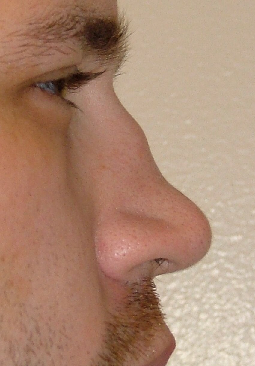
    WG's can present in many ways, but often involves a history or sinusitis or rhinitis, along with constitutional symptoms, hemoptysis, renal failure, and occassionally polyarthralgias. Patients can develop a "saddle nose" due to collapse of their nasal support.

    The pathological triad of WG includes:
    1) systemic necrotizing angiitis
    2) necrotizing granulomatous inflammation of the respiratory tract
    3) necrotizing glomerulonephritis

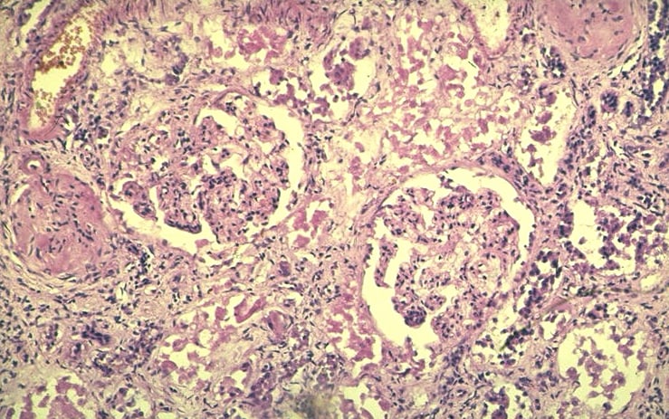
    Diagnosing Wegener's requires a detailed history and physical exam as well as blood work, looking for evidence of c-ANCA or anti-PR3, a urinalysis looking for RBC casts, chest imaging, and often a biopsy of either the kidney or lung. Lung biopsies frequently reveal evidence of vasculitis and granulomatous inflammation. Renal biopsies demonstrate a Pauci Immune picture (i.e.: no deposition of immunoglobulin or complement) with segmental crescentic necrotizing glomerulonephritis.

    Treatment includes pulsed steroids and often cyclophosphamide urgently as this is a rapidly progressive disease. Involvement of a rheumatologist and nephrologist for management and follow-up is usually warranted.

    Goodpasture's disease is a triad of pulmonary hemorrhage, glomerulonephritis (GN), and circulating anti-glomerular basement membrane (anti-GBM) antibodies in the blood. However, not all patients present with the triad, but anti-GBM antibodies is a distinguishing feature of Goodpasture's. Most patients (~60 - 80%) present with both pulmonary and renal disease, 20 -40% have only renal disease, and less than 10% have only pulmonary disease.

    Diagnosis, like WG, involves a thorough history and physical exam as well as blood work looking for anti-GBM antibodies, chest imaging, and usually a renal or lung biopsy. Direct immunofluorescence of the renal biopsy will light up and stain along the basement membrane, reflecting the deposition of IgG.

    Treatment again, requires rapid aggressive management; however, in the setting of Goodpasture's there is a role for plasmapheresis to remove any circulating anti-GBM antibodies. This occurs in conjunction with immunosuppression with pulsed steroids and often cyclophosphamide. Again, consultation with a nephrologist is often warranted!

    Here is a review on the differential diagnosis of hemoptysis and here is a brief case report and review of Goodpasture's from The Lancet.

    Tuesday, July 20, 2010

    Lymphadenopathy


    Today we discussed the unfortunate case of a 25 year old young man presenting with a multitude of complaints, including 4-weeks of right shoulder pain, 8-months of lower back pain, head ache, RUQ pain, dyspnea, non-productive cough, night sweats, and an "intentional" weight loss of ~30 pounds over 2 months. He had recently returned from South America, where he had spent the past few months.


    We generated a broad differential diagnosis for this diffuse constellation of complaints. The 3 main categories we reviewed were:

    1) Neoplasm - hematologic vs. solid organ (ie: testicular, given his gender and age)
    2) Infectious - TB, liver abscess, amoebic infection, liver fluke, malaria, infectious endocarditis
    3) Inflammatory - inflammatory bowel disease, SLE, sarcoidosis

    A CT scan revealed multiple lesions within the liver as well as porta hepatis and para-aortic lymphadenopathy. Large volume lymphadenopathy was also seen in the chest, along with some lytic bone lesions. The clinical picture, along with these imaging findings, raise the most likely diagnosis to lymphoma.

    Lymphomas are a heterogeneous group of cancers arising from the reticuloendothelial and lymphatic systems, which often present as solid tumors of lymphoid cells. Historically lymphomas have been categorized as Hodgkins Lymphoma (HL) or Non-Hodgkin Lymphoma (NHL), but as we learn more and more about these cancers the classifications are constantly being upgraded. Currently lymphomas are still broadly categorized as HL and NHL, with the latter group being further subclassified based on the type of cell from which they originate: B-cell, T-cell, or Natural Killer cell.

    HL results from clonal transformation of cells of B-cell origin, giving rise to the pathognomic Reed-Sternberg cells. The cause is unknown, but genetic susceptibility and environmental associations are thought to play a role. HL is slightly more common in patients with immunosuppression secondary to post-transplant drugs, congenital immunodeficiency, HIV, and autoimmune diseases (SLE, RA, Sjogren's, etc.). Patients often present with painless lymphadenopathy, that can become painful after consumption of alcohol... for no clear reason! Usually the lymphadenopathy arises in one area and spreads in a contiguous manner. The classic "B symptoms" are often described: fever, night sweats, and unintentional weight loss. Diagnosis requires a lymph node core biopsy +/- a fine needle biopsy. Depending on the specific type of HL identify on pathology, and the stage at diagnosis, treatment usually involves chemotherapy +/- radiation therapy, surgery, or stem cell transplant.

    NHL is also a disease of clonal transformation, most often of B-cells, but different subtypes of NHL can develop as a result of clonal transformation of T-cells or Natural Killer cells; classification depends on immunophenotyping, genotyping, and cytogenetics. NHL is much more common than HL. Immunosuppressed patients are also at risk for NHL. The exact etiology of NHL is yet to be discovered, but it is hypothesized that a viral cause may be at play. Patients often present again with painless lymphadenopathy; however, in NHL they can often have multiple areas of involvement. Occasionally patients can present with SVC syndrome secondary to compression of the SVC from nodal enlargement, or acute renal failure secondary to obstruction of the ureters from retroperitoneal or pelvic lymph nodes.

    Diagnosis requires a lymph node core biopsy for nodal architecture as well as immunophenotyping and cytogenetics for subclassification of the lymphoma. Full imaging with CT of the chest/abdo/pelvis and a BM biopsy is required for staging +/- a PET scan (this image shows lymphoma with increased uptake of 18-FDG in the brain, chest, and spleen). Although not fully validated in clinical settings, PET scans can be used in the proper setting, as determined by the oncologist and the radiologist.

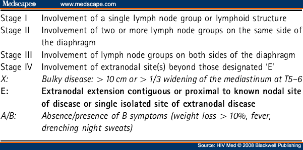
    Staging of NHL follows the Cotswold Modified Ann Arbour Staging. Prognosis is generally better for those with B-cell NHL compared to T-cell NHL. The International Prognostic Index (IPI) is used to help guide clinicians and patients with aggressive lymphomas. There are 5 risk factors with the IPI, and outcomes deteriorate as the number of risk factors increases.

    1) Age over 60

    2) Poor performance status measured by the ECOG (Eastern Cooperative Oncology Group)

    3) Elevated LDH

    4) More than 1 extranodal site

    5) Stage III or IV disease

    Patients in the highest IPI risk group (4-5 risk factors) have a 50% survival rate at 5-years, whereas those in the lowest IPI risk group (no risk factors) have a very high cure rate. Modifications to the IPI have been made to account for diffferent NHL, including the FLIPI (follicular lymphoma) and the R-IPI (diffuse large B-cell lymphoma).

    Treatment for NHL is again specific to the subtype of lymphoma and the stage at diagnosis; however, it generally incorporates chemotherapy "R-CHOP" (rituximab (anti-CD20 monoclonal antibodies), cyclophosphamide, doxorubicin, vincristine, prednisone), radiation therapy, or both +/- stem cell transplantation.

    Here is a great review article on the approach to a patient with lymphadenopathy.

    Friday, July 9, 2010

    Welcome to the Horses & Zebras of TGH

    Welcome to GIM at TGH and this, the official blog of the TGH CMR!

    This is my first posting and I will try to update the blog every few days with summaries from morning report, noon rounds, physical exam rounds, etc. as well as links to articles and web sites that provide excellent reviews of the topics at hand. Today's post will be a brief summary of the past week's salient teaching points!

    Last week we discussed an interesting case of seizure in a patient with a recent history of malaria, travel, and fever. We reviewed the general breakdown of the most common causes of seizure by age category:

    Children:
    -febrile seizure, seizure disorder
    Adults (~20 - 50):
    -trauma, toxic, alcohol withdrawal, metabolic, seizure disorder/idiopathic
    -less likely stroke or tumour
    Older Adults (>50):
    -more likely stroke or tumour
    -could still be one of the other causes


    Last Friday we discussed a great case of HCV cirrhosis and SBP, spontaneous bacterial peritonitis. Here is a NEJM review article on the management of cirrhosis and ascites. There is also a very helpful video on NEJM demonstrating how to perform a paracentesis found here. And this all links in well with Physical Exam Rounds yesterday, where we examined a patient with ascites. You should all have received the JAMA Rational Clinical Exam paper entitled "Does this patient have ascites?" in your inbox (it is not possible to link to the website!). Key points to remember for physical examination of ascites:
    1) Most sensitive findings are ANKLE EDEMA and INCREASED GIRTH
    2) Most specific finding is FLUID WAVE


    During our Emergency Lecture Series we have had a variety of excellent topics covered, including a review of atrial and ventricular arrhythmias. Here you will find the 2006 ACC/AHA guidelines for patients with Atrial Fibrillation as well as the 2003 guidelines for patients with SVT. Here you will find the ACC Pocket Guide for ventricular arrhythmias. And on that note, here is a link to drugs that can induce Torsades de Pointes.


    Today's Morning Report discussed a fascinating case of SOB, muscle weakness, swollen joints, and weight loss. The patient was found to have an elevated CK and a small pericardial efffusion on 2D Echo, but normal inflammatory markers (ESR and CRP). There were no sensory findings and only mild objective muscle weakness on exam.

    During our discussion the topic of Constrictive Pericarditis came up and how to make the diagnosis; which requires a right and left heart catheterization to compare pressures across the septum. Constrictive pericarditis is a relatively rare diagnosis that is often caused by tuberculosis or other infections agents such as fungi and parasites. Here is a great article, as mentioned in Morning Report, describing a case of constrictive pericarditis!

    For our case, there is no diagnosis as of yet - hopefully Team 5 will keep us all posted! However, as we discussed, our differential for this case includes (but is not limited to):

    1. Malignancy with associated Polymyositis/Dermatomyositis
    2. Rheumatoid arthritis
    3. Inclusion body polymyositis
    4. Mixed connective tissue disorder

    Dermatomyositis (DM) is less likely given that the patient has no skin manifestations. The classic skin features of DM include:

    Gottron's papules: lacy, pink/violaceous, raised or macular lesions, symmetric over the dorsal interphalangeal joints, elbows, and knees
    Heliotrope rash: a violaceous discolouration of the eyelids with periorbital edema
    Shawl sign/V-sign: erythematous rash over the shoulders/neck (in the pattern of a shawl) OR neck/chest (V-sign)
    Periungual telangiectasias: nail changes (cuticular hypertrophy) with or without Raynaud's
    Mechanic's hands: coarse, fissured, scaly, hyperkeratotic hands

    Thanks to former CMR David Frost for the DM links! For a complete review of DM please see this great review article in American Family Physician and a previous Blog by David Frost.

    Monday, June 28, 2010

    Signing off...


    This is the last blog post of the year for me...


    I've enjoyed managing the TWH Tangents and TGH Horses and Zebras Blogs for 2009-2010. I hope you found them helpful


    Your CMR 2009-2010

    Monday, June 14, 2010

    Pancytopenia


    Today we discussed an approach to the patient with pancytopenia

    This is an indication for a bone marrow aspirate and biopsy, which usually shows increased or decreased marrow cells:

    Hypocellular marrow
    -Marrow toxins: May be dose-dependent, predictable as with chemotherapeutic agents, immunomodulators (methotrexate, cyclophosphamide, etc) or idiosyncratic (chloramphenicol, sulfa drugs, phenytoin, many others); radiation, EtOH
    -Infection: Parvovirus B12, EBV, HIV, HHV-6, viral hepatitis; severe sepsis
    -Immune: SLE, RA (rare), graft-versus-host
    -PNH
    -Idiopathic stem cell failure

    Hypercellular marrow
    -MDS, leukemias, lymphomas, myeloma
    -Nutritional- severe megaloblastic anemia (B12, folate)
    -Marrow replacement/infiltration: myelophthisic anemia , myelofibrosis, granulomatous disease (TB, sarcoid, fungal), solid tumor metastases (prostate and breast are most common)

    The topic of the MGUS and multiple myeloma 'spectrum' came up. Here are some helpful criteria based on the NEJM review paper cited below:

    MGUS
    Marrow plasma cells less than 10% and M-protein (in blood) less than 30g/L
    No clinical manifestations

    Smouldering MM
    Marrow plasma cells at least 10% and/or M-protein at least 30g/L
    No clinical manifestations

    Multiple myeloma
    Marrow plasma cells over 10% and/or M-protein at least 30g/L
    Clinical features of hypercalcemia, renal failure, anemia, bone lesions, infections, plasmacytomas

    Waldenstrom's macroglobulinemia
    Marrow plasma cells over 10% and M-protein over 30 (IgM)
    Clinical features of anemia, bleeding, organomegaly, IgM immunoglobulin

    Primary amyloidosis (AL)
    Marrow plasma cells less than 10% and M-protein less than 30
    Clinical fearures of fatigue, wt loss, purpura, nephrotic syndrome, CHF, neuropathy, orthostatic hypotension, massive hepatomegaly

    Links:
    Click here for a "medical mystery" from NEJM on panyctopenia
    Click here for an excellent review paper on MGUS/myeloma differentiation

    Saturday, June 12, 2010

    Eating disorders- medical complications


    Today we discussed the medical complications of eating disorders, which can be devastating and affect any system, with mortality as high as 10% in anorexia and 2% in bulemia. A few points:


    Electrolyte/renal abnormalities
    -Hypokalemia (may be from vomiting, diuretic abuse, laxative abuse)
    -Metabolic alkalosis (volume depletion, diuretics)
    -Hyponatremia (diuretics, polydipsia prior to weighings)
    -Hypophosphatemia
    -Hypomagnesemia
    -Chronic kidney disease (may be contributed to by nephrocalcinosis, tubular dysfunction)

    Cardiovascular
    -Bradycardia (may or may not be secondary to electrolyte abnormalities)
    -Cardiomyopathy (potentially reversible)
    -Long QT
    -Mitral valve prolapse

    GI
    -Parotic hypertrophy
    -Decreased contractility
    -Mallory-Weiss teats
    -SMA syndrome
    -Gallstones

    Endocrine
    -Amenorrhea
    -'Sick euthyroid' syndrome
    -Growth retardation
    -Hypoglycemia

    Skeletal
    -Osteopenia/osteoporosis

    Hematologic
    -Marrow suppression

    Neurologic
    -Seizures
    -Peripheral neuropathy

    Dermatologic
    -Lanugo hair
    -Hair loss
    -Yellow/orange skin- hypercarotenema
    -Russell sign (callus over knuckles)
    -Edema

    Refeeding syndrome: When pts become rapidly "anabolic" after prolonged periods of starvation, severe electolyte abnormalities can develop and must be aggressively replaced.
    -Hypophosphatemia
    -Hypokalemia
    -Hypoglycemia
    -Edema

    Link:
    Click here for a NEJM review of anorexia
    Click here for a case report of reversible severe cardiomyopathy from anorexia

    Tuesday, June 8, 2010

    Status Epilepticus


    Today we discussed an approach to status epilepticus and seizures in general.

    Defined as continuous or repeated seizures without return to baseline for 20-30min (brain activity; may not have convulsions).
    NB- there is a movement to make the time part of this definition much shorter (to 5 minutes)

    Most seizures last 1-2 min. You need to intervene if beyond this point. At 20 min, the seizure per se damages the brain (scarring, self-perpetuating as sz focus). Also treat if series of short sz with incomplete recovery in between.

    Approach is to treat the seizure AND look for the underlying cause

    Acute causes: bleed, stroke, trauma, metabolic (e.g. hypoglycemia, hyponatremia, hypocalcemia, others), infections (esp. CNS), hypoxia, intoxication/drugs (or withdrawal), major organ faiure (uremia, hepatic failure)
    Chronic: mass, non-compliance with meds

    Complications of sz: aspiration, orthopedic complications, lactic acidosis, rhabdomyolysis

    Practical approach:

    ABC- clear the airway (do NOT put anything in the pt's mouth to prevent 'tongue biting, etc), roll on side, ensure breathing (apply O2)
    Get monitors- Vitals, O2 sat,
    IV access
    CHECK THE GLUCOSE!, If low, amp of D50, glucagon 1mg IM if no IV
    Thiamine 100mg IV (esp. if EtOH, pregnant, cancer pts)
    Blood: CBC, lytes, renal, Ca profile, glucose, transaminases, drug levels, tox screen

    If no immediately reversible cause (e.g. hypoglycemia), drugs to use acutely:

    Benzos- lorazepam IV 12-24h anti-sz effect. Loraz 0.1mg/kg at 1-2mg/min (usually 1-2mg aIV at a time, repeated to max) This will stop 80% of sz.
    May give diazepam PR if no IV access. 0.2mg/kg PR x 1 or midazolam 5mg IM x1

    Phenytoin (Dilantin) 20mg/kg IV load at 50mg/min (e.g. 1400mg over 30 min in 70kg)
    If needed, give further 5-10mg/kg.
    A couple of points about phenytoin for status:
    Do not mix phenytoin and D5; it crystallizes, which is not very helpful in status epilepticus.
    Never give dilantin and dopamine together; can cause profound hypotension for unclear reason

    Phenobarbital: Strongly consider intubation here
    20mg/kg IV @50-75mg/min. This pt should be in ICU

    Others: Propofol infusion, midazolam infusion

    Look for underlying- bloodwork, CT, LP if fever.
    Mimics of status: myoclonus, rigors, movement disorders, herniation (early)

    Link:

    Click here for an excellent review of status epilepticus from Chest (yes, a weird place for it to be)


    Thursday, June 3, 2010

    Endocarditis


    Today we discussed endocarditis. This is a very important topic to have knowledge of regardless of what area one practices in, both in terms of suspecting and confirming the diagnosis, and proper management. The natural history of untreated endocarditis is 100% mortality. A few points:

    Traditional breakdown is by time course; perhaps less relevant today than previously.

    Subacute classically presents as a chronic, wasting illness, with fever, wt loss and progressive heart failure and complications as the heart is progressively damaged over weeks to months. Typical organisms are (in order) viridans group streptococci, enterococcus, st. aureus

    Acute presents with rapidly progressive valvular destruction over days, often with catastrophic hemodynamic and other complications. Typical organisms are st. aureus, enterococci, viridans group streptococci, HACEK organisms.

    Other useful classifications that help with predicting microbiology and complications are native vs. prosthetic valve and R-sided vs. L-sided.

    Some physical findings (not exhaustive!)

    Signs of valvular involvement or local complications:
    -murmurs (esp. AI, MR, TR)
    -signs of L or R-sided heart failure
    -bradycardia (heart block can occur from paravalvular abscess)

    Signs of embolic complications/phenomena and vasculitis
    -any focal neurological deficit
    -peripheral cutaneous signs: Splinter hemorrhages, Osler nodes, Janeway lesions, Roth spots, clubbing, subconjuntival and other mucosal petechiae. Remember to look for these on the feet too
    -splenomegaly

    Signs of underlying causes
    -track marks for IDU
    -oral exam for dental hygiene

    Diagnosis:
    -multiple blood cultures from different sites off antibiotics are KEY to the diagnosis and management

    Modified Duke's criteria can be helpful (see link for full details):

    Major
    -multiple blood cultures consistent or Q-fever serology +ve
    -echo consistent (oscillating mass, abscess, dehiscence of prosthesis)
    -new murmur

    Minor
    -predisposing heart condition or IVDU
    -fever
    -vascular phenomena (emboli, etc as above)
    -immunologic phenomena (as above)
    -blood cultures not meeting criteria for "major"

    Definite: (2 major) or (1 major+3 minor) or (5 minor)
    Possible: (1 major and 1 minor) or (3 minor)

    ECG- check for new blocks (esp. 1st degree AVB; suggests abscess)

    Therapy
    General principles- need cidal antibiotic. Generally need minimum 4-6 wks of IV therapy

    Indications for valve replacement (NB- surgery is probably under-utilized)
    -CHF as a direct consequence
    -Severe valvular insufficiency
    -Paravalvular abscess
    -Embolic phenomena post-initiation of therapy
    -Persistently positive cultures or fever despite adequate medical therapy
    -Size of vegetation (relative)

    Links:
    Click here for the complete modified Duke's criteria
    Click here for a review article from NEJM

    Monday, May 31, 2010

    Paroxysmal Nocturnal Hemoglobinuria


    Today we discussed a very rare cause of anemia and thrombosis, PNH.

    This disease results from an acquired mutation in DNA encoding components of complement system, which leads to several clinical manifestations:

    1) Hemolysis, which is episodic ("paroxysmal"). The degree of hemolysis depends on how many RBCs are abnormal. Unclear why it is sometimes nocturnal, as shown above. Chronic hemolysis can cause severe anemia, and renal failure from myoglobin-induced tubular damage

    2) Thrombosis. It is unclear why patients with PNH are hypercoagulable; it may have to do with platelet abnormalities in PNH (all hematopoietic stem cells can be involved). Thrombosis is usually venous, but can be arterial. The hallmark is thromboses in relatively unusual places, most commonly portal vein or other intra-abdominal veins and cerebral venous sinus thrombosis.

    3) Diminished hematopoiesis. Because marrow stem cells are involved, pancytopenia can be ther presenting manifestation.

    Diagnosis: "PNH assay", which is flow cytometry for the clone of cells with abnormal complement-related proteins.

    Therapy:

    1) For hemolysis:
    -supportive measures include transfusion, iron supplementation, folic acid supplementation.
    -specific therapy is Eculizumab, a monoclonal antibody that inhibits complement activation, and significantly reduces hemolysis. Limiting factor is cost, estimated at $400,000 / year

    2) For thrombosis:
    -anticoagulation with heparin then warfarin as secondary prevention
    -there may be lower risks of thrombosis on Eculizumab
    -there is retrospective evidence supporting prophylactic anticoagulation with warfarin in selected patients

    Stem cell transplant is also an option reserved as a last resort

    Links:
    Click here for the paper showing the effect of Eculizumab in PNH

    Thursday, May 20, 2010

    Cocaine- medical complications


    Today we discussed cocaine intoxication. Some points:

    Acute intoxication:
    Cocaine is a sympathomimetic, and acute intoxication presents with this toxidrome:
    Hypertension, tachycardia, fever, flushing, diaphoresis, mydriasis, altered mental status, possibly seizures

    The anticholinergic toxidrome presents very similarly. The 2 can be distinguished by:
    -diaphoresis in sympathomimetic (vs. 'dry as a bone' in anticholinergic)
    -more prominent hypertension in sympathomimetic

    Management:
    Besides the specific organ issues discussed below, general managment consists of
    -benzodiazepines (mainstay!)
    -if BP control is required, DO NOT USE PURE BETA BLOCKERS; this causes unopposed alpha agonism and makes hypertension/vascular effects worse; labetalol is safe in this situation (because it is both alpha and beta-blocker), and phentolamine or phenoxybenzamine are alpha-blockers that can be used.

    Common specific organ effects (besides acute intoxication):
    CNS- cerebral vasoconstriction, including stroke; movement disorders
    CV- coronary vasospasm, which may cause MI; tachyarrhythmias, hypertensive urgency/emergency
    Resp- perforated septum, smoking cocaine causes SOB, wheezing, chest pain hemoptysis in a large proportion of users. Acute fever, dyspnea, hypoxia, infiltrates soon after smoking has been called "crack lung"; is from alveolar damage. Interstitial lung disease is possible.

    Recently recognized complication:
    In the past year or so, there have been many recognized cases (including at this hospital) of patients presenting with agranulocytosis from cocaine "contaminated" with levamisole, an anti-helminth drug used in animals.

    Click
    here for a summary of this complication
    Click here for a paper from the Annals of Internal Medicine describing this. CMAJ has a similar report.

    Wednesday, May 19, 2010

    Dilated cardiomyopathy


    Today we discussed causes of dilated cardiomyopathy,

    Some etiologies of DCM:

    1) Ischemia
    This is the most common cause of cardiomyopathy in the developed world. Within the category of "initially unexplained" DCM, ischemia is less common, since this is usually the first etiology excluded.

    2) Valvular heart disease
    Some valvular lesions are compensated chronically by LV dilation- mitral regurgitation and aortic insufficiency are the most common (although NB- MR can follow DCM from dilation itself; this is often termed "functional MR"). End-stage aortic stenosis can also cause DCM

    3) Infections
    -Viral infections can cause myocarditis, which can lead to DCM. Coxsackie, adeno, echo are the most common. HIV is a recognized as a cause of DCM
    -Chagas' disease (a protozoan infection) is most common cause of DCM in Central and South America.
    -Lyme disease is associated with cardiac muscle dysfunction, although heart blocks are more common.

    4) Toxins
    -Alcohol; amount and duration correlate with risk
    -Cocaine; may be independent of ischemia
    -Anthracycline-based chemotherapy (usually takes a dose of 450mg/M2; each cycle has about 50)
    -Trace elements. Rare cause; cobalt, arsenic, mercury... Click here for an interesting "outbreak" of cobalt-induced DCM in Quebec

    5) Genetic

    6) Peripartum CM- unclear cause; occurs in late pregnancy and early post-partum. Carries relatively good prognosis, but high risk of recurrence in subsequent pregnancies

    7) Infiltrative CM's- hemochromatosis, sarcoidosis, amyloidosis

    8) Tachycardia-induced cardiomyopathy. Prolonged tachycardia can cause reversible, sometimes severe, LV dysfunction

    9) "Stress cardiomyopathy" AKA "Takotsubo" cardiomyopathy:
    Rare, but increasingly recognized cause of LV dysfunction, with characteristic apical ballooning on 2D echo. May be associated with ECG changes including ST elevation. Sometimes associated with a stressful event (physiological or psychological); self-resolving. Click
    here for the origin of this term, the Japanese octopus trap...

    In a significant proportion of cases, the cause remains idiopathic.

    Links:
    Click here for a recent NEJM review of DCM that goes over the evidence base for various pharmacologic and device therapies

    Click here for a study looking at underlying causes and prognosis for initially idiopathic dilated cardiomyopathy.


    Friday, May 14, 2010

    Hyperthyroidism- physical exam


    Today we discussed some of the physical exam findings of hyperthyroidism and how to diagnose a goiter

    Hyperthyroidism
    A general approach to the examination for hyperthyroidsm with some possible findings
    General appearance: restless, anxious, thin
    Vitals- tachycardia, atrial fibrillation, wide pulse pressure, fever
    Eyes-
    lid lag (superior sclera visible as patient looks down),
    lid retraction, widened palpebral fissure
    Graves' orbitopathy (see below)
    CNS- fine tremor with hands outstretched, increased reflexes, proximal muscle weakness
    CV- flow murmur
    Abdo- increased bowel sounds
    Extremities- warm, moist skin, velvety skin, pretibial myxedema (in Graves)
    Associations: other autoimmune- e.g. vitiligo, adrenal insufficiency

    A word about eye findings in hyperthyroidism
    Some eye findings are general to all causes of hyperthyroidism (from increased sympathetic activity), and some are specific to Graves' disease.

    Sympathetic
    -lid lag
    -lid retraction
    -stare
    -conjunctival injection

    Graves orbitopathy
    -EOM involvement
    -lid edema
    -exophthalmos
    -loss of visual acuity

    Evidence for how to diagnose a goiter:
    Inspect and palpate
    -look for lateral prominence = distance from imaginary line between cricoid and suprasternal notch to surface of thyroid gland
    -Categorize as normal or goiter
    -Subcategorize as small (1-2x) or large (over 2x) goiter

    Ruling out a goiter:
    Normal size by palpation (negative R 0.15)
    No lateral prominence (NLR 0.41)
    “Goiter ruled out” = normal thyroid size or gland not visible with neck fully extended

    Ruling in a goiter:
    Estimated size by inspection;/palpation
    1-2x normal (positive LR 1.9)
    over 2x normal (PLR 25)
    Lateral prominence 0-2mm (PLR 3.4)
    Lateral prominence > 2mm (PLR infinity)
    “Goiter ruled in” = large goiter or lateral prominence over 2mm

    Link:

    Click here for link to JAMA Rational Clinical Examination for goiter (unfortunately, a special JAMA subscription is required)


    Wednesday, May 12, 2010

    Scleroderma


    Today we discussed scleroderma (AKA systemic sclerosis)

    This is a multisystem disesase of small vessels and skin/organ fibrosis

    Epidemiology: Age 35-65, Female more common than male (7-12:1).

    There are 2 types of systemic sclerosis- limited and diffuse

    Limited SSc: sclerosis, thickening of distal limbs without truncal involvement. CREST fits into this category. Associated with anti-centromere AB. Calcinosis refers to localized hand masses on fingers/forearms. Telangiectasias are seen in face/mucosa/hands. Most serious manifestations of limited are pulm HTN, digital ischemia.

    Diffuse SSc: thickening over distal and proximal limbs and trunk with significant organ involvement. Criteria are thickening of extremities proximal to MCPs or digital ischemia, pulmonary fibrosis. Edematous skin, painful joints, tendon friction rub, rapidly progressive skin fibrosis. Associated with SCL70 antibody.

    Clinical:
    Initially nonspecific; Raynaud's, fatigue, MSK complaints followed in variable time period by further sx (soon in diffuse, later in limited).
    First specific manifestations are skin thickening, starting as swelling/puffiness over fingers/hands.
    Subsequently variable; skin, pulmonary, cardiac, renal, GI involvement.

    90% of SSc pts have Raynaud's and fibrosis of fingers, with loss of digital pads (sclerodactyly) and digital ulceration.

    Skin: edema, tightness, decreased flexibility, contractures, then fibrosis.

    MSK: arthralgias, arthritis, calcinosis, tendon involvement. Late: weakness/atrophy, possible overlap myosits

    Pulm: Pulmonary fibrosis, PFTs show decreased volumes, diffusion impairment.

    GI: small mouth, dysphagia, GERD/esophagitis. Loss of peristalsis

    Cardiac: Myocardial fibrosis, myocarditis, diastolic dysfunction

    Renal: Pathological change in most (small vessel vasculopathy); extreme is scleroderma renal crisis (acute kidney injury, with microangiopathic hemolytic anemia)

    Therapy: No proven disease modifying agents; treatment is by organ system involved. In severe cases, immunosuppression may be used (e.g. steroids, methotrexate, cyclophosphamide)

    Skin- key is moisturize, treat pruritus
    Raynaud's- CCB, prazosin in severe
    GI- treat GERD, prokinetics for paresis
    Renal- ACE-I
    Pulmonary- measures for pulm HTN

    Links:
    Click here for a recent NEJM review of scleroderma
    Click here for a review from Rheumatology on scleroderma

    Monday, May 10, 2010

    Alcoholic hepatitis


    Today we discussed alcoholic hepatitis.

    This is distinct from chronic alcohol-induced liver disease, which causes steatosis and predisposes to fibrosis, then cirrhosis.

    Alcoholic hepatitis is a clinical syndrome of jaundice and liver failure (usually after decades of over 100g/d intake). Patients have often stopped drinking for several weeks before onset. Female sex is a risk factor for this specific condition (with equal intake to men), but more men have the typical intake required.

    Rapid onset of jaundice is the cardinal sign. Others include fever and ascites. Liver is typically enlarged and tender. Signs of chronic liver disease are often present.

    Alcoholic hepatitis is a poor prognostic indicator; 40% of patients with severe alcoholic hepatitis die within 6 months

    Diagnosis:
    Lab: AST usually above 2x ULN, but rarely over 1000. ALT is lower. WBC count (esp. neutrophils), and INR are usually high. Renal failure (if it is hepatorenal syndrome) carries a poor prognosis.
    Biopsy shows swollen hepatocytes with Mallory bodies. Cholestasis may be present.

    Ddx includes NASH, viral hepatitis, drug-induced, fulminant Wilson's, autoimmune liver disaese, alpha-1, pyogenic abscess, cholangitis, HCC.

    Severity indices:
    Maddrey's discriminant function: [4.6 x (pt's PTT - control PTT)] + bili (mg/dL)
    Value over 32 indicates severe alcoholic hepatitis; this is threshold for starting steroids.
    MELD over 21- severe

    Therapy:
    General
    salt restriction, diuretics, lactulose, thiamine, withdrawal tx as indicated. Tap for SBP
    EtOH abstinence
    Specific
    Steroids: prednisolone 40mg/d x 4 weeks, then taper (or equivalent)
    Pentoxyfylline- 400mg PO TID increases in-hospital survival; little head-to head data comparing to steroids; should be considered where there are contraindications to steroids.

    Link:
    Click here for a 2009 review from NEJM on alcoholic hepatitis that goes over the evidence for various therapies.

    Wednesday, April 28, 2010

    Syphilis


    Today we discussed syphilis. Some points about this infection that has made a resurgence:

    Treponema pallidum (spirochete), is spread through contact with infectious lesions or fluids. 50-75% of exposed sex partners get infected.

    Clinical manifestations (NB- syphilis is the "great imitator"; these are the more common presentation, but keep a high index of suspicion in the right epidemiological setting)

    Primary:
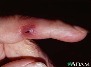
    -skin lesion (chancre) at site of innoculaion ~21d after exposure. Often goes unnoticed. Classically non-tender, indurated, non-purulent. Heals spontaneously.


    Secondary:
    -4-10 weeks after chancre appearance. Rash in 90%; pink, red, or purpule macules on flanks, shoulders, arms, chest, back. May become maculosquamous/papular and affect hands/soles in 50-80%.
    -other signs/symptoms include sore throat, malaise, h/a, lymphadenopathy. Can cause neurologic, renal, ophthalmologic, GI, hepatic disease. Resolves without treatment, may recur.

    Tertiary:
    Years later- aortitis, gumma (mass), iritis.

    A word about neurosyphilis:
    Neurosyphillis can occur early or late. May coexist with primary, secondary, or tertiary. May see meningitis, cranial nerve involvement, eye involvement, meningoencephalitis, meningovascular (presents as stroke). Late findings are rare; occur decades later: paresis, dementia with psychosis (rapidly progressive), tabes dorsalis (posterior column involvement, bowel, bladder dysfunction).

    LP indicated in any of 1) neuro/ocular/otic symptoms/signs. 2) biochemical evidence of late latent syphilis (or unknown duration) in HIV pt. 3) active tertiary- gumma, aortitis, iritis. 4) treatment failure for non-neurosyphilis

    Lab testing
    The lab testing for syphilis is very complex and confusing. Here are some general comments; click
    here for detailed interpretation from the Ontario Public Health Lab website

    NB- RPR is essentially synonymous with VDRL
    1) EIA (enzyme immunoassay), TPPA, FTA-Abs. All of these are durable antibody tests that are very sensitive, and remain positive for life. If negative, syphilis is usually ruled out. If positive, need confirmatory testing (there are false positives)
    2) RPR- This is also an antibody test, but more suggestive of active infection. If postive, this confirms diagnosis. If negative, it can mean 1 of several things: a) very early primary- retest in 2 weeks. b) successful treatment. c) late latent d) biologic false positive

    Treatment:
    NB- latent =seroreactivity without clinical manifestations. Early is infected within previous year by documented conversion. Late is over 1 year from primary infection

    Primary, secondary, early latent: Pen G 2.4M U IM single dose
    Late latent, unknown, tertiary: Pen G 2.4M U IM qweekly x 3.
    Neurosyphilis, ocular, otic: Pen G IV 18-24M units/d divided q4h for 10-14d.

    NB- need repeat serology to confirm/document treatment success- see paper below for details

    Primary should have RPR negative at 1 year. Secondary at 2 years. Tertiary at 5 years.
    Links:

    Click here for a JAMA review of syphilis that most of this post is based on
    Click here for the Ontario Public Health Lab algorithm for interpreting the MANY permutations and combinations of lab results

    Wednesday, April 21, 2010

    Myelopathy causes


    Today we discussed myelopathy (i.e. spinal cord pathology)

    Features to suggest myelopathy:
    -bilateral motor and sensory signs or symptoms not involving the head
    -weakness, spasticity, hyperreflexia (although may see hyporeflexia acutely)
    -sensory findings with a descrete level
    -bowel and bladder involvement

    Causes of myelopathy:

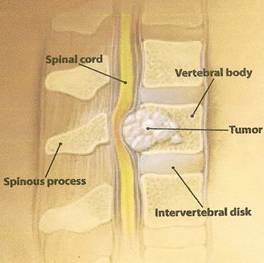
    1) Compression- herniated disc, tumor (extramedullary or intra-medullary) most common. Epidural abscess (often st. aureus). Also consider in conditions with c-spine instability like Down's and rheumatoid arthrititis

    2) Inflammatory- MS; typically incomplete. SLE- usually incomplete, within a vascular territory (i.e. transverse myelitis), CNS vasculitis

    3) Metabolic- subacute combined degeneration (i.e. dorsal column and corticospinal tract)- ddx is B12 deficiency and tabes dorsalis (syphilis)

    4) Infectious- cord itself: syphilis, HIV, HTLV (esp in Caribbean), VZV. Surrounding structures causing compression: epidural abscess, vertebral osteomyelitis (bacterial, mycobacterial or fungal)

    5) Vascular- cord infarction- rare (can be seen in aortic dissection or hypercoagulable state), AVM- very rare.

    6) Genetic- Devic's (optic neuritis, myelopathy only- probably variant of MS)

    Link:
    Click here for a NEJM review paper by UHN physicians on cervical radiculopathy and myelopathy