Monday, February 1, 2010

Nephrotic syndrome


Triad of hypoalbuminemia, edema, nephrotic-range proteinuria (over 3.5g/24h)
Other components of the syndrome: hyperlipidemia, hypercoagulability, lipiduria (causing oval fat bodies seen above)

The primary problem in the nephrotic syndrome is urinary protein loss due to altered permeability of the glomerulus. Other manifestations are all secondary to protein loss.

Loss of albumin and consequent loss of oncotic pressure lead to the clinical manifestation of edema, which may be profound. Periorbital edema is sometimes more prominent in the nephrotic syndrome than other causes of edema.

Hyperlipidemia results from urinary loss of regulatory lipoproteins, and low oncotic pressure may increase hepatic lipogenesis

Hypercoagulability may be arterial or venous, and often involves the kidneys themselves (i.e. renal vein thrombosis). May result from urinary loss of proteins c and s, and possibly antithrombin III

Causes in adults:

Primary renal diseases:
Minimal change
Focal segmental glomerulosclerosis (FSGS)
Collapsing glomerulopathy
Membranous nephropathy
Membranoproliferative GN (MPGN)
IgA nephropathy

Systemic diseases:
Infectious: HBV, HCV, HIV, syphillis, schistosomiaisis, malaria
Inflammatory/rheumatologic: SLE, amyloidosis, cryoglobulinemia
Malignancy: solid tumors, primary hematologic malignancies (e.g. Hodgkin's lymphoma)
Others: Sickle cell disease, heroin, drugs (e.g. gold, penicillamine)

Therapy:
1) treat the underlying cause
2) measures to limit proteinuria- largely ACE-inhibitors and/or ARBs
3) measures to deal with complications- salt restriction, careful diuretic use (as may become easily intravascularly depleted), statins, vitamin D supplementation if deficient

Links:

Click here for a NEJM clinical case on nephrotic syndrome


Tuesday, January 26, 2010

Fulminant hepatic failure


Defined as rapid onset of encephalopathy and marked decrease in liver synthetic function within 28d of symptom onset in patient without chronic liver disease

Major etiologies are 1)acetaminophen OD, 2) idiosyncratic drug reaction, 3) Hep B, 4) Hep A, 5) others

Coagulopathy usually precedes encephalopathy, which may quickly progress to coma

It is important to determine the etiology, since specific causes may have specific treatments:
NAC for acetaminophen, penicillin for amanita mushroom poisoning, delivery for acute fatty liver of pregnancy, zinc/trientine for Wilson's.

Pts should be admitted to ICU and transferred to a transplant centre.

Supportive care includes:
glucose (may need d10 drip)
electrolyte replacement
fluids
reversal of coagulopathy
neurochecks >q6h

FFP (4u) may normalize coags for 6h (for procedure or bleeding)

Empiric abx for any sign of infection

Poor prognostic indicators predicting need for transplant:
pH below 7.3 after resuscitation
PTT over 100
Cr over 290
Gr 3 or 4 encephalopathy
Lactate over 3.3 4h post admission


Major causes of transaminases in the thousands:

1) Ischemia
-inflow (hypotension- shock liver, PV thrombosis)
-outflow (CHF, Budd-Chiari)

2) Viral
HAV, HBV (rarely HCV)
Others- EBV, HSV, CMV

3) Drugs/toxins

4) Autoimmune

5) Obstruction/stone

6) Acetaminophen and EtOH both (but rarely EtOH alone)


Link:
Click here for a NEJM case of fulminant hepatic failure from HSV infection that discusses management considerations
Click here for a paper on approach to increased liver enzymes

Friday, January 22, 2010

Celiac disease


Today we discussed malabsorption, and one of its causes, celiac disease. A few key points:

Clinical features of malabsorption:
Chronic diarrhea (esp. steatorrhea)
Weight loss despite adequate intake
Deficiency of fat-soluble vitamins: (ADEK); vit D deficiency may lead to hypocalcemia, vit K deficiency may lead to high INR
Hypocalcemia
B12 and folate deficiency
Iron deficiency
Hypoalbuminemia

Celiac disease is an autoimmune disease involving the small bowel, precipitated by intake of gluten. This autoimmune reaction leads to chronic small bowel inflammation.

Long term complications include small bowel adenocarcinoma and small bowel T-cell lymphoma

Celiac was traditionally thought of as a malabsorption syndrome of children, but it is increasingly recognized in adults, even the elderly. A common way patients come to clinical attention is iron deficiency anemia that does not respond to oral iron supplementation (because the etiology is malobsorption).
Diagnosis:
Gold standard for diagnosis is small bowel biopsy showing villous atrophy.

Serologic tests are also available that have different performance characteristics:
Endomesial IgA antibodies are near 100% specific (remember to check IgA levels, as many patients with celiac disease have low IgA levels)
Tissue transglutaminase antibodies have sensitivity of over 90%, and this is the screening test
Gliadin antibodes are less sensitive and specific (~80% Sn and Sp)

Therapy:
Gluten-free diet
Treat nutritional deficiencies
Celiac disease is associated with hyposplenism; patients should receive the pneumococcal vaccine
Bone loss is common; patients should have a DEXA scan and treatment as appropriate

Monitoring:
Symptoms should improve with gluten-free diet
Serology can be used to monitor response; antibody titers correspond to degree of inflammation.
Non-response should raise suspicion of small bowel lymphoma or non-adherence to gluten-free diet

Link:
Click here for a NEJM review on celiac disease

Monday, January 18, 2010

Acetaminophen toxicity


Today we discussed acetaminophen toxicity. Some key points:

Stages of toxicity:
1) preclinical toxic effect with normal ALT (may see no elevation for first 24h)
2) hepatic injury (elevated ALT)
3) hepatic failure (injury with hepatic encephalopathy)- pts presenting here have 20-40% mortality

Glutathione is necessary for the pathway to a non-toxic metabolite. When it is depleted, NAPQI (the toxic metabolite) is formed.

Overdoses (acute or chronic) deplete glutathione and allow liver injury. N-acetylcysteine (NAC) restores glutathione.

If within 2h of ingestion, activated charcoal is used. Beyond 2h, benefit is limited. With a known time of ingestion, the
Rumack-Matthew nomogram is used; if level is above line for time of ingestion, there is risk of hepatic injury and NAC should be given.

When no time is known, usual practice is to treat anyone who may have overdosed and has measurable level.

The nomogram is not useful with chronic ingestion, unknown time of ingestion, and with long-acting preparations

There is a PO NAC protocol (72h) and an IV NAC protocol (20h). There are no head to head trials comparing.

Most toxicologists recommend checking acetaminophen level and ALT prior to stopping NAC. Criteria for stopping NAC are falling ALT and undetectable acetaminophen level.

High risk features (i.e. predictive of fulminant failure): encephalopathy, INR over 6.5, Cr over 300, pH below 7.3, hypoglycemia, hyperbilirubinemia. Note that AST/ALT levels are not predictive of poor outcome; only indicate injury.

Link:
Click here for a NEJM review of acetaminophen toxicity

Friday, January 15, 2010

Stroke in the young patient


Today we discussed stroke in a young patient. Most references use under 45 as the cutoff for invoking some of the possibilities discussed below.

Differential diagnosis to consider for any patient presenting with "stroke", particularly a young patient:

Structural disease (tumor, bleed), seizure (Todd's paresis), dissection, migraine (esp. migraine sensory aura), demyelination, hypoglycemia. Anything that can cause delirium can cause focal findings when superimposed on a structurally abnormal brain.

In pts 45 and younger, common causes of ischemic stroke include
-Cardioembolic is #1 etiology (structural heart disease, endocarditis, others)
-Always consider dissection, even in the absence of trauma (esp if pain with onset). Dissections are usually extracranial, assoc with trauma or neck manipulation.
-Vasculitis
-Hypercoagulable state
-Cocaine, amphetamines
-Sickle cell disease
-Migranous stroke
More exotic causes: CADASIL, MELAS, Moya-moya

In many patients, an underlying cause is never found.
Workup:
CT/MRI/MRA/MRV
TEE
carotid doppler
thrombophilia screen
skin bx if suspect CADASIL
Antiphospholipid antibodies

Link:
Click here for NEJM review of PFO and stroke in young patients

Tuesday, January 12, 2010

NMS and serotonin syndrome


Today we discussed neuroleptic malignant syndrome and serotonin syndrome. A few points:

Neuroleptic malignant syndrome:
Syndrome of altered mental status, fever (often over 40), marked rigidity, CK elevation (usually over 1000), and autonomic instability (including hypertension, arrhythmias, tachycardia).

Seen in the context of

1) New or altered dose of antipsychotic medication (usually "typicals")
2) Withdrawal of dopaminergic medications (e.g. antiparkinsonians)
Treatment is discontinuation of drug (or restarting if withdrawal), possibly dantrolene or bromocriptine)

There is an extensive differential diagnosis of components of this presentation, the most important of which to exclude are CNS infections (meningitis, encephalitis), other systemic infections, seizures, intoxications, withdrawal states.

Serotonin syndrome
Related, but distinct disorder that results from overstimulation of serotonin receptors.

It occurs in the setting of substances that either increase serotonin release (e.g. ecstasy, cocaine, amphetamines), or inhibit serotonin reuptake (SSRIs, SNRIs, tricyclics, MAOIs)

Presentation can include mental status change, diarrhea, flushing, diaphoresis, tremor, restlessness.
Physical exam may show fever, hyperreflexia, myoclonus, rigidity, autonomic instability.

Tx: Stop serotonergic substance. If severe, aggressive cooling, benzos, cyproheptadine (antihistamine with antiserotonin effect)

NMS vs. serotonin syndrome differences
Serotonin syndrome may have shivering, hyperreflexia, myoclonus, flushing (all uncommon in NMS)

NMS tends to have more prominent CK elevation, rigidity and higher temperatures

Poison control consultation is an important component of managing both of these situations.

Links
Click here for a CMAJ review of NMS
Click here for a CMAJ review of serotonin syndrome

Monday, January 11, 2010

Acute visual loss


Today we discussed the concerning symptom of acute visual loss. Some points:

Monocular visual loss:

1) TIA (i.e. amaurosis fugax; curtain-like classically) - if not transient, represents branch or central retinal artery occlusion. These pts are at high risk for stroke. May see cherry red fovea and Hollenhorst plaques (cholesterol)

2) Central retinal vein (or branch) occlusion- can be transient or permanent, sometimes associated with hypercoagulability. May see engorged veins, hemorrhage.

3) Anterior Ischemic Optic Neuropathy (AION).
This is divided into "arteritic" and "non-arteritic".
Arteritic essentially means temporal arteritis.
Non-arteritic means no specific cause; HTN and DM2 are risk factors

4) Optic neuritis- demyelination, compression (abscess, tumor), hereditary causes

5) Primary ophthalmologic causes: Retinal detachment, vitreous hemorrhage or detachment, closed angle glaucoma, maculopathy,

Acute binocular blindness suggests a toxic cause (e.g. methanol, carbon monoxide, ethabutol, others). Stroke (cortical blindness from visual cortex involvement) and migraine are also on the differential.

Link:
Click
here for a BMJ website outlining approach to visual loss

Tuesday, August 4, 2009

Non-Resolving Pneumonia

Factors to Consider in Non-Resolving Pneumonia

(1) Do not confuse "non-resolving pneumonia" with non-resolution of chest x-ray findings. Although radiographic resolution has been used in the past to define this entity, Mittl and colleagues demonstrated that only half of patients with community acquired pneumonia have radiographic resolution at two weeks. Clearance was faster in non-smokers and those treated as outpatients. Having said that, patients with radiographic evidence of pneumonia do require follow-up imaging to ensure resolution and to rule out an underlying mass lesion.

(2) The definition endorsed by the Infectious Disease Society of North America is fairly vague- "a situation in which an inadequate clinical response in present despite antibiotic treatment". As in many things, clinical judgment is paramount - consider ongoing cough with sputum production, fever, performance status, hypoxia and white blood cell count.

(3) The possible etiologies are many and include both infectious and non-infectious causes. Important things to consider are:
  • Time of treatment - Patients treated less than 72 hrs should be considered as inadequate treatment time.
  • Infectious Causes - Consider a pathogen not covered by your treatment (e.g. tuberculosis, non-tuberculous mycobateria, viral or fungal infections) or a resistant organism (e.g. MRSA pneumonia).
  • Non-Infectious Causes - malignancy, interstitial lung disease, heart failure. (The NEJM recently published a report of a patient with assymetric pulmonary edema due to mitral valve dysfunction).
  • Complications of Infection - Empyema or parapneumonic effusion.
(4) Approach to Management
  • Does your patient need ISOLATION? Patients with non-resolving pneumonia should be considered high-risk for tuberculous and influenzae, depending on their epidemiology.
  • Does your patient need urgent antimicrobial treatment? If not, consider stopping all antimicrobials to increase the yield of investigations.

  • Investigations for everyone - blood cultures
  • Investigations to consider - HIV serology, induced sputum, bronchoscopy, CT chest. Very rarely, a patient may need a lung biopsy to make a diagnosis.
  • Patients with a pleural effusion need diagnostic thoracentesis, and chest tube insertion if an empyema is identified.

For the full IDSA guidelines on community acquired pneumonia, including a review of the approach to a non-resolving pneumonia, visit the IDSA website.

Wednesday, July 29, 2009

Salicylate toxicity

Salicylate toxicity is not a common phenomenon in internal medicine, but it is important to recognize it because the management is specific.

A couple of general principles to remember in overdoses and toxidromes:

(1) Assess ABCs early and frequently. Patients can change.
(2) There is no such thing as too much IV access. Patients with toxic ingestions can seize, become hypotensive and do other unpredictable things. You will never be sorry that you asked for two IVs.
(3) Always call POISON CONTROL.
(4) Consider multiple ingestions.
(5) Think about general principles of overdose management, including decontamination.
(6) The patient may need additional monitoring, possibly in the intensive care unit setting. This is particularly true if the level of consciousness if depressed or if there is significant hypotension. Again, remember that patients change and a patient who does not need ICU at one moment may later on.

Salicylate toxicity comes in two forms:
(1) Acute overdoses - often intentional, with self-harm of suicidal intent.
(2) Chronic overdoses - from regular high dose ASA use.

The presentation can be variable and non-specific, although there are some cardinal features.

  • Tachycardia, hyperventilation, fever
  • Altered level of consciousness - related to direct toxicity, hypoglycemia, cerebral edema.
  • Nausea, vomiting
  • Platelet dysfunction
  • Pulmonary edema (especially with chronic toxicity, more common in the elderly)
  • Tinnitus (especially with chronic toxicity)

**The combination of an anion gap metabolic acidosis and a primary respiratory alkalosis (beyond compensation for the metabolic acidosis) should trigger the diagnosis of salicylate toxicity**

Specific Management Guidelines
(1) Alkalanize the urine. This is done by running a sodium bicarbonate infusion, targeting a urine pH of 7.5-8.5. In general, start the infusion at 150-200mL/hr and increase according to your urine pH.
(2) Potassium replacement. For any patient with urine output and a serum potassium <5, consider replacement. Similarly to patients with diabetic ketoacidosis, the total body potassium may be low despite a normal measured serum potassium because of intracellular shift related to acidosis. With alkalanization, you may see a precipitous drop in the serum K.
(3) Nephrology consultation +/- hemodialysis - Consider in patients with severe toxicity, acute renal failure, pulmonary edema. If you are not sure, involve nephrology.
(4) Consider ICU consultation. Consider endotracheal intubation if necessary from the perspective of airway protection. However, mechanical ventilation may not be as effective as the patient's own respiratory drive in correcting their metabolic acidosis.

Monday, July 20, 2009

Bacterial Meningitis

Today we discussed the approach to the diagnosis and management of bacterial meningitis.


For a good general review of bacterial meningitis, read the NEJM review by van de Beek et al. The Infectious Diseases Society of America also provides Clinical Practice Guidelines.

I've focused the discussion here on a few evidence based points that were discussed this morning.

(1) Pre-treatment with antibiotics prior to lumbar puncture may cause the culture to become negative, but should not change the biochemical properties of the CSF. This was shown in a study published by Schaad et al comparing ceftriaxone to cefuroxime where repeat lumbar punctures were done at 24 hours post initiation of antibiotics and were found to be unchanged in terms of WBC count, protein and glucose. Based partly on this data, guidelines recommend that if the patient requires a CT scan prior to LP, antibiotics should be given after blood cultures have been drawn, but prior to CT or lumbar puncture.

(2) Concurrent administration of dexamethasone with or prior to the first dose of antibiotics reduces mortality. This difference was shown in a 2002 study published in the NEJM. In this study of a combined group of patients with both streptococcus pneumoniae and with neisseria meningitidis, although the difference was largely evident in the s. pneumoniae group. The dose of dexamethasone used was 10 mg Q6H and this is the current standard of care.

(3) As shown in this brief report, re-insertion of the stylet post lumbar puncture decreases the risk of post-LP headache.

(4) Which patients need a CT brain prior to lumbar puncture? Although the vast majority of CTs done in these patients are normal, the IDSA guidelines recommend CT for anyone with an altered LOC, focal neurologic deficits, papilledema, an immunocompromised state, history of CNS disease or new onset seizure. I've linked the NEJM article supporting these guidelines here.

Saturday, July 18, 2009

Physical Exam - Aortic Stenosis

To determine if a systolic murmur is related to aortic stenosis, consider the following predictive factors:

The following factors are SENSITIVE (help to rule OUT aortic stenosis)
No systolic murmur
No radiation of murmur to right clavicle

The following are SPECIFIC (help to rule IN aortic stenosis)
Pulsus parvus
Plusus tardus
Decreased S2
Brachioradial delay
Apical carotid delay
Mid-late peaking murmur

Two very good evidence based evaluations supporting this are the JAMA Rational Clinical Exam Series paper on systolic murmurs and the JGIM paper on aortic stenosis. Both come from Toronto clinicians, and are linked here and here.

Monday, July 13, 2009

Mesothelioma


Today we discussed the evaluation of a new pleural effusion in the context of a history of smoking and possible occupational asbestos exposure. The approach to pleural effusions is reviewed here.

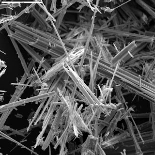
One interesting disease that was brought up today was malignant mesothelioma. Although most commonly found as a pleural based malignancy, mesothelioma is probably best described as a cancer of serosal surfaces and can rarely present as peritoneal disease with ascites or pericardial disease.

The incidence of malignant mesothelioma is expected to rise until at least 2020, an epidemiologic phenomenon that lags behind known exposure to asbestos. In fact, in parts of the developing world, asbestos is an ongoing exposure, and therefore mesothelioma may be an ongoing problem for many years.

The diagnosis of mesothelioma can be difficult, and the sensitivity of cytology of pleural or ascitic fluid is quite variable, ranging from 33-84%, often necessitating needle or thorascopic biopsy. The disease usually presents at an advanced stage when large effusions have reached the point of symptoms, and median survival is less than one year from the time of diagnosis. Management is typically palliative, and may include local management such as pleurodesis or chronic drainage, or systemic therapy. Occasionally surgery is required to manage complications of locally advanced disease.

A good NEJM review is available.

Tuesday, July 7, 2009

Welcome to "The Pulse", a blog of educational activities at TGH. This blog was started by Isaac Bogoch last year and will be continued onwards this year. All errors, omissions and mis-representations are his.

Today at morning report, we discussed the approach to a patient with an altered level of consciousness. The facilitator provided a framework for the differential diagnosis:

1. Neurologic causes - including stroke, CNS infections, dementing illnesses such as Lewy Body dementia, normal pressure hydrocephalus and others.

2. Metabolic causes - including electrolyte abnormalities (think sodium and calcium!), endocrinopathies (e.g. thyroid disease).

3. Medications - overdose, intoxication, withdrawal. Also consider over-the-counter and alternative therapies.

4. Major medical illness. Both liver and kidney dysfunction can present with altered level of consciousness.

5. Psychiatric illness. Depression can present in atypical ways, particularly in the elderly.

We also talked about the basics of the workup and management of stroke. A good general review article on stroke can be found in the New England Journal of Medicine 2007, Vol 357 (6), pp 572-579. A link to the abstract is found here

I would encourage you to consult the Canadian Stroke Network best practice guidelines on stroke management, linked here. Although practice guidelines often represent a combination of evidence and expert opinion, they can be helpful resources and can direct you to the peer-reviewed literature.

Hope you are all enjoying the first week. Let me know how I can help make this a great experience for you!

Shannon

Wednesday, June 24, 2009

Over-and-out


This will be the last Morning Report Blog update of the academic year. I hope this was a valuable learning experience for you. It's been a good time.

Your CMR, 2008/2009

Rhabdomyolysis

Rhabdomyolysis....

First, think about the underlying etiology:

1. Trauma: this can be overt, like a crush injury or a bit more subtle, like immobilization in an elderly person who falls and is unable to get up for some time. Also think about immobilization in patients with a decreased level of consciousness or during prolonged operation.

2. Physical activity: rhabdo may occur in those who either perform excessive physical activity (eg. marathons), in those who are doing significantly more physical activity than they are used to (eg. couch potato who goes on run for 1st time in 10 years), or in situations where hyperthermia may occur (eg. jogging in the Sahara). Also, don't forget seizures as a common etiology.

3. Drugs/Toxins: as always, we should consider prescribed drugs (eg statins, colchicine), and non-prescribed drugs (eg. alcohol, cocaine, ecstasy). There are always a few cases per year of rhabdomyolysis from wild mushroom poisoning.

4. Infections: many viruses (eg. cytomegalovirus, Coxsackievirus, Epstein-Barr, Influenza, adenovirus, HIV), bacteria (eg. pyomyositis), sepsis, and parasitic (Falciparum malaria).

5. Electrolyte: primarily hypokalemia and hypophosphatemia from any cause.

6. Endocrinopathy: mostly in hypothyroidism, but may also be seen in DKA/HONK - probably from hypophosphatemia.

7. Those who are more prone: people who have myopathies may be more prone to developing rhabomyolysis. Think about those with poly/dermatomyositis, malignant hyperthermia, or rare congenital myopathies.

Other: paraneoplastic syndromes

What should I watch out for?

  1. Hyperkalemia: lots of potassium can be released from muscle cells. Monitor this and the ECGs closely.
  2. Renal failure: watch out. Myoglobin is toxic to the renal tubules and can cause acute tubular necrosis.
  3. Other electrolytes: hyperphosphatemia (released from muscle cells), hypocalcemia.


Treatment:

This primarily revolves around finding and reversing precipitants, and aggressive fluid administration to prevent myoglobin-induced ATN. There is debate whether the best fluids are saline or if sodium bicarbonate added to D5W works best. Also, keep a close eye on the potassium.

A good link:

A strange case of rhabdomyolysis from CMAJ.


Tuesday, June 23, 2009

Malnutrition


Evaluation for Malnutrition:
  • General Inspection: look for grooming, BMI <19, cachexia
  • Vital signs: increased HR if intravascularly volume depleted
  • Head & Neck: alopecia and brittle hair, glossitis (Iron, B12), bleeding gums (Vit C), angular cheilitis (Iron)
  • Abdominal Exam: look for ascites, hepatomegaly, splenomegaly
  • MSK: Muscle wasting. Pay particular attention to temporal muscle wasting, deltoids - note a 'boxing off' appearance, triceps, and quadriceps muscles. Also assess if ribs can be visualized through pectoral muscles. Note subcutaneous fat loss in these areas as well.
  • Neuro: look for loss of deep tendon reflexes, peripheral neuropathy, optic atrophy, and evidence of subacute combined degeneration of the cord (B12).

A Rational Clinical Exam article from
JAMA was published by local talent explains how to determine if your patient is malnourished via the Subjective Global Assessment. See below

Thursday, June 18, 2009

"Elite" or "Advanced" Morning Report


Today we had a Morning Report session with our friends from Mt. Sinai and Toronto Western Hospitals. We discussed a very interesting case of a person with shortness of breath in the context of rheumatoid arthritis.

  • Please see the Mt. Sinai Blog for details here.
  • More on interstitial lung disease can be found here.

Friday, June 5, 2009

A total overreaction

(the syndrome of Reactive arthritis pictured left)

Reactive Arthritis
- (formerly known as Reiter's Syndrome)

What is it? A post infectious immunologic phenomenon resulting in arthritis.

How long after an infection does it develop? a few days to a few weeks. This lag-time may make it difficult to know what the offending pathogen was.

Which pathogens are associated? Think about two big systems....GI and GU
  • GI: Yersinia, Salmonella, Shigella, Campylobacter
  • GU: Chlamydia, and possibly Gonorrhea

What does the syndrome look like?
The arthritis is usually an asymmetric oligoarthritis,. Enthesitis is common and may affect the achilles tendon or plantar fascia on the calcaneus. Conjunctivitis or uveitis is seen, as is balanitis - Hence the adage "Can't pee, can't see, can't climb a tree". A classic dematologic manifestation of reactive arthritis is keratoderma blennorrhagica - scaly lesions on the palms and soles. See picture below.

Is there any genetic predisposition to developing Reactive Arthritis? Yes.

What is it? HLA-B27 is present in just under 50%.

How do we treat it? NSAIDs work well, like indomethacin or naproxen. In severe cases, DMARDs like sulfasalazine may be used.


Why is it called Reactive Arthritis now instead of Reiter's Syndrome? an interesting paper can be found here.

(keratoderma blennorrhagica pictured left)

Wednesday, June 3, 2009

Measles

Measles

No, this is not just an infection of children anymore - we are seeing measles more frequently in adults. This is usually in those with no history of primary vaccination.

Classic Measles: there is about a 14 day incubation period where the virus replicates and spreads via lymphatics and hematogenously. Initial symptoms include fever, coryza, conjunctivitis, cough, and general malaise. Koplik's spots may be seen as well - these are small, raised lesions in the buccal mucosa that are whitish or blue in colour (see photo below). These are pathognomonic for measles and can be visualized roughly 2 days prior to the classic rash.

The exanthem of measles typically starts on the face and moves to the trunk and extremities, sparing the hands. It is a blanchable maculopapular rash (see photo above). Patients may start to feel better a couple of days after the rash appears, and the rash typically will start to fade after 4ish days.

Diagnosis: can be confirmed with serology. Send off measles IgM - this should be positive a few days after the exanthem appears. Measles IgG will be detectable two weeks after the exanthem.

What to do? this is a very contagious virus, so patients with suspected measles should be in respiratory isolation and the Infection Control service should be contacted. Treatment revolves mostly around supportive care - fluids, antipyretics, and monitoring for/treating bacterial suprainfections like pneumonia or otitis media. Vitamin A is given to children with measles in countries where vitamin A deficiency is prevalent.

Links:
  • A neat case and image of Koplik's spots can be found here.
  • Check out the epidemiology of measles in North America here.

(Koplik's Spots in the buccal mucosa)

Tuesday, June 2, 2009

Pericarditis...


Today we discussed prognostic tools for pneumonia - the PORT and CURB65 scores. You can read more about these here.


As part of our differential diagnosis of Chest Pain, the topic of pericarditis came up.

Clinical presentation: usually a sudden onset of retrosternal chest pain with a pleuritic component to it, often relieved by sitting up. You may hear a pericardial rub - this is classically described as a triphasic, high-pitched sound. The 'tri' refers to:
  1. atrial systole
  2. ventricular systole
  3. ventricular diastole

It may be a transient phenomenon so listen again if you don't hear it. Classic situation...you hear the pericardial rub, admit and treat the patient. When your attending reviews the case the next morning they can't hear it - even though you swear on your life that you heard one. Solution: when you hear a pericardial rub, get your friend to have a listen as well, so in the morning both of you can say you heard it.

ECG: may show diffuse, concave ST elevations that do not fit any particular vascular territory. PR depression is also seen. Check out the ECG above.

Treatment: In most cases of idiopathic pericarditis, high dose NSAIDS are effective. Steroids and colchicine also may have a role. Interestingly, newer evidence suggests that colchicine may be a good first line agent. Here is a link to the article published in Circulation. Have a look and decide for yourself.

Other Links: