Wednesday, May 19, 2010

Dilated cardiomyopathy


Today we discussed causes of dilated cardiomyopathy,

Some etiologies of DCM:

1) Ischemia
This is the most common cause of cardiomyopathy in the developed world. Within the category of "initially unexplained" DCM, ischemia is less common, since this is usually the first etiology excluded.

2) Valvular heart disease
Some valvular lesions are compensated chronically by LV dilation- mitral regurgitation and aortic insufficiency are the most common (although NB- MR can follow DCM from dilation itself; this is often termed "functional MR"). End-stage aortic stenosis can also cause DCM

3) Infections
-Viral infections can cause myocarditis, which can lead to DCM. Coxsackie, adeno, echo are the most common. HIV is a recognized as a cause of DCM
-Chagas' disease (a protozoan infection) is most common cause of DCM in Central and South America.
-Lyme disease is associated with cardiac muscle dysfunction, although heart blocks are more common.

4) Toxins
-Alcohol; amount and duration correlate with risk
-Cocaine; may be independent of ischemia
-Anthracycline-based chemotherapy (usually takes a dose of 450mg/M2; each cycle has about 50)
-Trace elements. Rare cause; cobalt, arsenic, mercury... Click here for an interesting "outbreak" of cobalt-induced DCM in Quebec

5) Genetic

6) Peripartum CM- unclear cause; occurs in late pregnancy and early post-partum. Carries relatively good prognosis, but high risk of recurrence in subsequent pregnancies

7) Infiltrative CM's- hemochromatosis, sarcoidosis, amyloidosis

8) Tachycardia-induced cardiomyopathy. Prolonged tachycardia can cause reversible, sometimes severe, LV dysfunction

9) "Stress cardiomyopathy" AKA "Takotsubo" cardiomyopathy:
Rare, but increasingly recognized cause of LV dysfunction, with characteristic apical ballooning on 2D echo. May be associated with ECG changes including ST elevation. Sometimes associated with a stressful event (physiological or psychological); self-resolving. Click
here for the origin of this term, the Japanese octopus trap...

In a significant proportion of cases, the cause remains idiopathic.

Links:
Click here for a recent NEJM review of DCM that goes over the evidence base for various pharmacologic and device therapies

Click here for a study looking at underlying causes and prognosis for initially idiopathic dilated cardiomyopathy.


Friday, May 14, 2010

Hyperthyroidism- physical exam


Today we discussed some of the physical exam findings of hyperthyroidism and how to diagnose a goiter

Hyperthyroidism
A general approach to the examination for hyperthyroidsm with some possible findings
General appearance: restless, anxious, thin
Vitals- tachycardia, atrial fibrillation, wide pulse pressure, fever
Eyes-
lid lag (superior sclera visible as patient looks down),
lid retraction, widened palpebral fissure
Graves' orbitopathy (see below)
CNS- fine tremor with hands outstretched, increased reflexes, proximal muscle weakness
CV- flow murmur
Abdo- increased bowel sounds
Extremities- warm, moist skin, velvety skin, pretibial myxedema (in Graves)
Associations: other autoimmune- e.g. vitiligo, adrenal insufficiency

A word about eye findings in hyperthyroidism
Some eye findings are general to all causes of hyperthyroidism (from increased sympathetic activity), and some are specific to Graves' disease.

Sympathetic
-lid lag
-lid retraction
-stare
-conjunctival injection

Graves orbitopathy
-EOM involvement
-lid edema
-exophthalmos
-loss of visual acuity

Evidence for how to diagnose a goiter:
Inspect and palpate
-look for lateral prominence = distance from imaginary line between cricoid and suprasternal notch to surface of thyroid gland
-Categorize as normal or goiter
-Subcategorize as small (1-2x) or large (over 2x) goiter

Ruling out a goiter:
Normal size by palpation (negative R 0.15)
No lateral prominence (NLR 0.41)
“Goiter ruled out” = normal thyroid size or gland not visible with neck fully extended

Ruling in a goiter:
Estimated size by inspection;/palpation
1-2x normal (positive LR 1.9)
over 2x normal (PLR 25)
Lateral prominence 0-2mm (PLR 3.4)
Lateral prominence > 2mm (PLR infinity)
“Goiter ruled in” = large goiter or lateral prominence over 2mm

Link:

Click here for link to JAMA Rational Clinical Examination for goiter (unfortunately, a special JAMA subscription is required)


Wednesday, May 12, 2010

Scleroderma


Today we discussed scleroderma (AKA systemic sclerosis)

This is a multisystem disesase of small vessels and skin/organ fibrosis

Epidemiology: Age 35-65, Female more common than male (7-12:1).

There are 2 types of systemic sclerosis- limited and diffuse

Limited SSc: sclerosis, thickening of distal limbs without truncal involvement. CREST fits into this category. Associated with anti-centromere AB. Calcinosis refers to localized hand masses on fingers/forearms. Telangiectasias are seen in face/mucosa/hands. Most serious manifestations of limited are pulm HTN, digital ischemia.

Diffuse SSc: thickening over distal and proximal limbs and trunk with significant organ involvement. Criteria are thickening of extremities proximal to MCPs or digital ischemia, pulmonary fibrosis. Edematous skin, painful joints, tendon friction rub, rapidly progressive skin fibrosis. Associated with SCL70 antibody.

Clinical:
Initially nonspecific; Raynaud's, fatigue, MSK complaints followed in variable time period by further sx (soon in diffuse, later in limited).
First specific manifestations are skin thickening, starting as swelling/puffiness over fingers/hands.
Subsequently variable; skin, pulmonary, cardiac, renal, GI involvement.

90% of SSc pts have Raynaud's and fibrosis of fingers, with loss of digital pads (sclerodactyly) and digital ulceration.

Skin: edema, tightness, decreased flexibility, contractures, then fibrosis.

MSK: arthralgias, arthritis, calcinosis, tendon involvement. Late: weakness/atrophy, possible overlap myosits

Pulm: Pulmonary fibrosis, PFTs show decreased volumes, diffusion impairment.

GI: small mouth, dysphagia, GERD/esophagitis. Loss of peristalsis

Cardiac: Myocardial fibrosis, myocarditis, diastolic dysfunction

Renal: Pathological change in most (small vessel vasculopathy); extreme is scleroderma renal crisis (acute kidney injury, with microangiopathic hemolytic anemia)

Therapy: No proven disease modifying agents; treatment is by organ system involved. In severe cases, immunosuppression may be used (e.g. steroids, methotrexate, cyclophosphamide)

Skin- key is moisturize, treat pruritus
Raynaud's- CCB, prazosin in severe
GI- treat GERD, prokinetics for paresis
Renal- ACE-I
Pulmonary- measures for pulm HTN

Links:
Click here for a recent NEJM review of scleroderma
Click here for a review from Rheumatology on scleroderma

Monday, May 10, 2010

Alcoholic hepatitis


Today we discussed alcoholic hepatitis.

This is distinct from chronic alcohol-induced liver disease, which causes steatosis and predisposes to fibrosis, then cirrhosis.

Alcoholic hepatitis is a clinical syndrome of jaundice and liver failure (usually after decades of over 100g/d intake). Patients have often stopped drinking for several weeks before onset. Female sex is a risk factor for this specific condition (with equal intake to men), but more men have the typical intake required.

Rapid onset of jaundice is the cardinal sign. Others include fever and ascites. Liver is typically enlarged and tender. Signs of chronic liver disease are often present.

Alcoholic hepatitis is a poor prognostic indicator; 40% of patients with severe alcoholic hepatitis die within 6 months

Diagnosis:
Lab: AST usually above 2x ULN, but rarely over 1000. ALT is lower. WBC count (esp. neutrophils), and INR are usually high. Renal failure (if it is hepatorenal syndrome) carries a poor prognosis.
Biopsy shows swollen hepatocytes with Mallory bodies. Cholestasis may be present.

Ddx includes NASH, viral hepatitis, drug-induced, fulminant Wilson's, autoimmune liver disaese, alpha-1, pyogenic abscess, cholangitis, HCC.

Severity indices:
Maddrey's discriminant function: [4.6 x (pt's PTT - control PTT)] + bili (mg/dL)
Value over 32 indicates severe alcoholic hepatitis; this is threshold for starting steroids.
MELD over 21- severe

Therapy:
General
salt restriction, diuretics, lactulose, thiamine, withdrawal tx as indicated. Tap for SBP
EtOH abstinence
Specific
Steroids: prednisolone 40mg/d x 4 weeks, then taper (or equivalent)
Pentoxyfylline- 400mg PO TID increases in-hospital survival; little head-to head data comparing to steroids; should be considered where there are contraindications to steroids.

Link:
Click here for a 2009 review from NEJM on alcoholic hepatitis that goes over the evidence for various therapies.

Wednesday, April 28, 2010

Syphilis


Today we discussed syphilis. Some points about this infection that has made a resurgence:

Treponema pallidum (spirochete), is spread through contact with infectious lesions or fluids. 50-75% of exposed sex partners get infected.

Clinical manifestations (NB- syphilis is the "great imitator"; these are the more common presentation, but keep a high index of suspicion in the right epidemiological setting)

Primary:
-skin lesion (chancre) at site of innoculaion ~21d after exposure. Often goes unnoticed. Classically non-tender, indurated, non-purulent. Heals spontaneously.


Secondary:
-4-10 weeks after chancre appearance. Rash in 90%; pink, red, or purpule macules on flanks, shoulders, arms, chest, back. May become maculosquamous/papular and affect hands/soles in 50-80%.
-other signs/symptoms include sore throat, malaise, h/a, lymphadenopathy. Can cause neurologic, renal, ophthalmologic, GI, hepatic disease. Resolves without treatment, may recur.

Tertiary:
Years later- aortitis, gumma (mass), iritis.

A word about neurosyphilis:
Neurosyphillis can occur early or late. May coexist with primary, secondary, or tertiary. May see meningitis, cranial nerve involvement, eye involvement, meningoencephalitis, meningovascular (presents as stroke). Late findings are rare; occur decades later: paresis, dementia with psychosis (rapidly progressive), tabes dorsalis (posterior column involvement, bowel, bladder dysfunction).

LP indicated in any of 1) neuro/ocular/otic symptoms/signs. 2) biochemical evidence of late latent syphilis (or unknown duration) in HIV pt. 3) active tertiary- gumma, aortitis, iritis. 4) treatment failure for non-neurosyphilis

Lab testing
The lab testing for syphilis is very complex and confusing. Here are some general comments; click
here for detailed interpretation from the Ontario Public Health Lab website

NB- RPR is essentially synonymous with VDRL
1) EIA (enzyme immunoassay), TPPA, FTA-Abs. All of these are durable antibody tests that are very sensitive, and remain positive for life. If negative, syphilis is usually ruled out. If positive, need confirmatory testing (there are false positives)
2) RPR- This is also an antibody test, but more suggestive of active infection. If postive, this confirms diagnosis. If negative, it can mean 1 of several things: a) very early primary- retest in 2 weeks. b) successful treatment. c) late latent d) biologic false positive

Treatment:
NB- latent =seroreactivity without clinical manifestations. Early is infected within previous year by documented conversion. Late is over 1 year from primary infection

Primary, secondary, early latent: Pen G 2.4M U IM single dose
Late latent, unknown, tertiary: Pen G 2.4M U IM qweekly x 3.
Neurosyphilis, ocular, otic: Pen G IV 18-24M units/d divided q4h for 10-14d.

NB- need repeat serology to confirm/document treatment success- see paper below for details

Primary should have RPR negative at 1 year. Secondary at 2 years. Tertiary at 5 years.

Links:

Click here for a JAMA review of syphilis that most of this post is based on
Click here for the Ontario Public Health Lab algorithm for interpreting the MANY permutations and combinations of lab results

Wednesday, April 21, 2010

Myelopathy causes


Today we discussed myelopathy (i.e. spinal cord pathology)

Features to suggest myelopathy:
-bilateral motor and sensory signs or symptoms not involving the head
-weakness, spasticity, hyperreflexia (although may see hyporeflexia acutely)
-sensory findings with a descrete level
-bowel and bladder involvement

Causes of myelopathy:

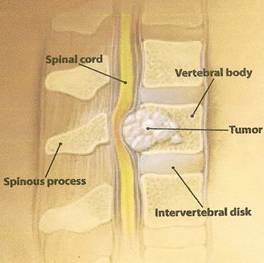
1) Compression- herniated disc, tumor (extramedullary or intra-medullary) most common. Epidural abscess (often st. aureus). Also consider in conditions with c-spine instability like Down's and rheumatoid arthrititis

2) Inflammatory- MS; typically incomplete. SLE- usually incomplete, within a vascular territory (i.e. transverse myelitis), CNS vasculitis

3) Metabolic- subacute combined degeneration (i.e. dorsal column and corticospinal tract)- ddx is B12 deficiency and tabes dorsalis (syphilis)

4) Infectious- cord itself: syphilis, HIV, HTLV (esp in Caribbean), VZV. Surrounding structures causing compression: epidural abscess, vertebral osteomyelitis (bacterial, mycobacterial or fungal)

5) Vascular- cord infarction- rare (can be seen in aortic dissection or hypercoagulable state), AVM- very rare.

6) Genetic- Devic's (optic neuritis, myelopathy only- probably variant of MS)

Link:
Click here for a NEJM review paper by UHN physicians on cervical radiculopathy and myelopathy

Thursday, April 15, 2010

Dyspnea in a patient with cancer


The general approach to the patient with a malignancy who presents with dyspnea is similar to that in a patient without a malignancy, but some considerations are more prominent in this situation. Some issues to think about:

Infections:
-post-obstructive pneumonia
-opportunistics: PCP, especially if on prednisone 20mg for over 2 months (or equivalent).
-nosocomial pneumonia
-fungal esp. if neutropenia.
-TB reactivation
-tachypnea as compensation for lactic acidosis in sepsis

Vascular/lymphatic:
-pulmonary embolism
-lymphangitic carcinomatosis
-tumor emboli syndrome (seen in lung, prostate, breast- micromets plugging pulmonary capillaries. PFTs may show diffusion abn).
-secondary pulmonary HTN

Airway:
-endobronchial lesion
-COPD- esp smoker with lung ca,

Neuromuscular:
-Lambert-Eaton
-phrenic nerve involvement
-steroid myopathy

Parenchymal/interstitial (in addition to infectious items above):
-drug effect
-radiation effect

Pleural:
-pleural effusion
-pneumothorax
-pleural involvement of malignancy itself

Cardiac:
-pumonary edema seconary to cardiomyopathy- chemotherapy toxicity (esp. anthracyclines) -pericardial effusion

Other:
-anemia

Wednesday, April 14, 2010

CLL and its complications


Today we discussed CLL and some issues related to hyperleukocytosis

Chronic lymphocytic leukemia:
Briefly, the most common leukemia; commonly an indolent condition for up to decades, but a subset transform to another lymphoproleferative disorder (e.g. lymphoma) or AML.

Most commonly picked up as incidental finding of lymphocytosis >5000, splenomegaly, lymphadenopathy, smudge cells on blood film (shown above). Sometimes B symptoms. Is diagnosed by flow cytometry showing monoclonal B markers (bone marrow bx not always needed).

Complications of CLL:
1) Immunodeficiency as outlined below
2) Autoimmune hemolytic anemia

3) Red cell aplasia
4) Transformation as above

Immunodeficiency in CLL:
This is B-cell disorder, so humoral immunodeficiency.
This type of immunocompromise is seen in congenital hypogammaglobulinemia (common variable immunodeficiency), B cell disorders (inc. myeloma, Waldenstrom's, CLL). Causes susceptibility to encapsulated (s. pneumo, H. flu, meningococcus), as well as Giardia, and C. Diff.
IVIG may be indicated in severe infection or immunoglobulin level <0.5 lower limit of normal (see link below). These pts get recurrent sinopulmonary infections because they do not produce secretory IgA.

In patients with CLL who are treated, there can be severe immunodeficiency of other types:
1) Neutropenia from myelotoxic chemotherapy (e.g. chlorambucil)
2) Cell-mediated- from fludarabine (t-cell inhibitor sometimes used); susceptible to PCP, listeriosis, fungal, etc.

This makes a severe, combined immunodeficiency state (like congenital SCID)

Leukostasis:
More important if cells are sticky (esp blasts). Causes leaky endothelium. May cause noncardiogenic pulm edema, retinal hemorrhage, encephalopathy, stroke, renal failure, MI, heart failure.
Not all leukocytosis is the same; risk of hyperviscosity/leukostasis depends on how 'sticky' the involved cell is:
Highest risk is blasts in AML.
If AML, very high risk is WBC over 100
CML- WBC in 200's or blast conversion
CLL-very rare to have leukostasis complications regardless of level.

If acute complications of leukostasis, hematologists/oncologists will sometimes use hydroxyurea, cyclophosphamide, or even leukopheresis

Link:

Click
here for the abstract of the RCT showing benefit of IVIG in pts with CLL with hypogammaglobulinemia and recurrent infections

Tuesday, April 13, 2010

Amyloidosis


Today we discussed amyloidosis. A few points about this rare, but serious and often multi-systemic condition

Amyloidosis refers to deposition of an altered conformation of a protein into amyloid fibrils that deposit systemically, giving various clinical manifestations depending on which system(s) are involved. One way of classifying amyloidosis is by which protein is forming the amyloid:

AL: amyloid is formed by immunoglobulin light chains or fragment produced by clone of plasma cells. Plasma cell burden is usually low (~5-10%). 10% have multiple myeloma.

AA: amyloid is formed by 'amyloid A protein', an acute phase reactant. This form of amyloid is associated with longstanding inflammatory conditions. Classic ones are RA, IBD, FMF

Familial amyloidosis: proteins forming amyloid are transthyreitin, apolipoproteins, lysozyme, etc.

"Senile systemic": transthyreitin amyloid, predominantly in heart.

Others: Alzheimer's disease is associated with amyloid fibril deposition in the brain; the protein is a-beta 40 and 42. It is unclear whether amyloid fibrils are causitive.

Possible clinical manifestations:
1) Neurological- CNS- intracranial hemorrhage (amyloid angiopathy), dementia. PNS- peripheral neuropathy (glove-stocking), compressive neuropathy (e.g. carpal tunnel)

2) Cardiac- infiltrative cardiomyopathy- systolic or diastolic dysfunction, arrhyhtmia (heart blocks or ventricular arrhythmias). Cardiac involvement carries the worst prognosis. Rare in AA amyloidosis.

3) Renal- may cause nephrotic syndrome or asynptomatic proteinuria

4) GI- hepatomegaly with or without splenomegaly, GI bleeding, dysphagia

5) MSK- macroglossia as shown above, arthritis

6) Dermatologic- ecchymoses from friable vessels,
periorbital purpura, fat infiltration is common but asymptomatic- may allow for diagnosis

7) Hematologic- Factor X deficiency leading to bleeding diathesis, petechiae (F10 binds to amyloid fibrils)

8) Pulmonary- less common; persistent pleural effusions

Diagnosis is made by organ system involved. Abdominal fat pad bx (stained for Congo red) is positive in 80% of AL, 65% of AA.

Treatment depends on type.
In AA, directed at underlying inflammatory disorder
In AL, directed at underlying plasma cell disorder

For cardiac amyloidosis, implanted defibrillator is sometimes indicated. High dose systemic chemotherapy (e.g. melphalan) is sometimes used.

Link:
Click here for a NEJM case discussion of amyloidosis- nicely summarizes the issues

Friday, April 9, 2010

Hypercalcemia


Today we had some discussion about hypercalcemia. Some points:

Symptoms:
GI- anorexia, n/v, abdo pain, constipation.
Renal: stones, polyuria.
Neuro: weakness.
Cardiac: arrhythmias

Etiology:
Useful first division is by PTH level
1) High PTH- expect high Ca, low PO4.
-primary/secondary/teriary hyperparathydoidism, parathyroid hyperplasia
-lithium
-familial hypercalcemic hypocalciuria

2) Low PTH
-PTHrP from malignancy (esp. SCC- lung, H+N)
-hypervitaminosis D- expect high Ca and high PO4- from granulomatous disease, lymphoma)C) -OAF = IL6 (local paracrine effect; in breast and hematological cancers)
-Direct effect of mets (e.g. prostate, lung, etc.)
-Myeloma
-Medications- HCTZ, Ca, vit. D
-Milk alkali syndrome
-Hyperthyroidism

As inpatient, #1 cause = malignancy
As outpatient, #1 cause = primary hyperparathyrodisim

Tx:
Fluids!
Consider bisphosphonate if malignancy-related or v. high (but takes days to work)
Calcitonin by nasal spray or subq.
If hyper D from sarcoid or lymphoma, possible steroids.
Avoid lasix since most patients are profoundly volume depleted.
Last resort is dialysis

Milk-Alkali syndrome
Triad of hypercalcemia, met alk, renal failure assoc with ingestion of large amts calcium, alkali
Once common because of PUD treatement. Making resurgence b/c of calcium for osteoporosis, and prevention of secondary hyperparathyroidism in CKD
Sequence: hypercalcemia, dec GFR, met alkalosis ("contraction"). Hypercalcemia per se stimulates renal bicarb fomation.
Pts on vit D, thiazides, vol contraction, CKD are at higher risk

Some links:
Click here for NEJM clinical problem solving case on hypercalcemia
Click here for a review of calcium disorders in renal disease

Tuesday, April 6, 2010

Atrial fibrillation- acute management


Today we discussed issues related to acute management of atrial fibrillation. A few points:

After establishing that the patient's airway and breathing are stable, the first decision point is
whether the patient needs emergent synched cardioversion.
Indications as per ACLS algorithm:
1) Hemodynamic instability, 2) Shock (i.e. hypoperfusion), 3) Pulmonary edema, 4) Ischemia
HOWEVER, remember that clinical judgement is absolutely required; this is not risk-free, and many patients have mild pulmonary edema and borderline hypotension with rapid A-fib and do not all need to be cardioverted.

If emergent cardioversion is indicated, important steps include
1) Consider pre-treatment with antiarrhythmic (e.g. amiodarone); increases the likelihood of success
2) Analgesia and sedation, requiring the capability to deal with the airway; usually requires the help of anesthesia unless in the ICU
3) Application of the pads- "sandwich"- front and back is more successful than sternal and lateral
4) Synchronized cardioversion with 50-100J
5) Anticoagulation for at least 4 weeks post-cardioversion

Watch for
-bradycardia (pts usually have some AV nodal agent "on board" prior that was dosed to their a-fib; if now in sinus, may be bradycardic
-embolic complications

If elective cardioversion (i.e. not emergent), considerations include
1) Embolic risk
If a-fib is present more than 48h (usually difficult to determine) or the patient was not therapeutically anticoagulated for 2 weeks or more prior, there is a significant (but difficult to quantify) risk of systemic embolization with restoration of sinus rhythm. A trans-esophageal echo that shows no evidence of a thrombus makes the risk very low, and is done in situations where emergent cardioversion is not necessary, and the above criteria are not met.
NB- the risk of embolization exists for pharmacologic cardioversion as well, although may be lower than electrical (think about this when using antiarrhythmics like amiodarone)

2) Procedural risk (sedation, analgesia, arrhythmia, etc)

If cardioversion is not indicated, rate control is the next management choice. Options include:
1) Beta-blockers (e.g. metoprolol 2.5-5mg IV over 5-10min)
2) Non-DHP calcium channel blockers (e.g. diltiazem 15-20mg IV over 5-10 min)
3) Digoxin (e.g. 0.25mg IV load)
4) Amiodarone (but note cardioversion risk as outlined above)

There is little evidence to guide us in the acute setting (but lots of evidence in the chronic setting- e.g. AFFIRM)
Reasons to use beta blockers or CCB: Little toxicity, easy to titrate, often the patient has another indication (for B-blocker), rate control is more reliable than digoxin (see below)
Reasons to think twice before using a beta blocker or CCB: LV dysfunction (esp. CCB), pulmonary edema (both), reactive airways (B-blocker), conduction system disease (both)

Reasons to consider digoxin: Is an inotrope, and carries lower risk of hypotension or worsening pulmonary edema. Works particularly well in decompensated aortic stenosis since allows more filling time without affecting contractility
Reasons to think twice before using digoxin: Patient can override it with sympathetic stimulation (that is usually present) since it does not block this, risk of toxicity if levels supratherapeutic, more difficult to use, debatable long-term benefit (definitely no mortality benefit from DIG trial)

Anticoagulation may be initiated in the ED if clearly indicated, but may also wait until the patient is more stable, and risks/benefits (which usually favour anticoagulation) have been discussed.

Monday, April 5, 2010

Lymphadenopathy


Today we discussed an approach to lymphadenopathy. A few points:

Normally, nodes are smaller than 1cm, mobile, soft, nontender. May be larger in young pts. Palpable inguinal nodes or submandibular can be normal.

Generally subdivide into local (1 grp) vs. general.
Local-

Cervical:
Infection- EBV, CMV, toxo, TB (lymphadenitis if intact, "scrofula" if burst), cat scratch (bartonella), staph, strep infections
Malignancy- lymphoma, metastatic squamous ca (esp. posterior)
Other- Kikuchi's disease (cervical LA and fever in young female)

Supraclavicular:
Malignancy- 24-50% of pts over 40 with supraclavicular adenopathy. R-sided is associated with thoracic (lung, mediastinum, esophagus). L-sided is assocaited with abdominal (virchow's node; especially gastric ca)

Axillary:
Infection- cat scratch, trauma
Malig- BrCa, mets

Epitrochlear:
Infection- local trauma, secondary syphilis, tularemia
Malig- lymphoma
Other- sarcoid

Inguinal:
Infection- lower ext/gu inf
malig- lymphoma

Generalized:

Malignancy- lymphoproliferative, leukemia, mets
Infection- HIV (acute seroconv), EBV (fever, lymphad, pharyngitis; post>ant), typhoid, CMV, histoplasmosis, mycobacterial infection (TB, MAI), others...
SLE
Serum sickness (=tetrad of fever, arthralgias, rash, LA). Drugs often cause: allopurinol, phenytoin, penicillin, hydralazine, carbamazepine, gold, etc)
Others: Sarcoidosis, Castleman's disease

Approach:

H: infection/malig, time course, pain, etc, exposures (bites, uncooked meat, sexual hx, drugs)
P: location, size, tenderness, consistency, mobility, spleen, look for primary

Inv: in generalized, CBC, lytes, ca, LDH, uric acid, PBF, CXR, HIV, ANA, VDRL. Localized: if no suggestion of malig, may observe for 3-4 wks. If persistent, biopsy.

Link:

Click here for an excellent article on lymphadenopathy from American Family Physician

Wednesday, March 31, 2010

Diplopia and dysconjugate gaze, ptosis


Today we discussed an approach to ptosis, diplopia and dysconjugate gaze. A few points:

An approach to diplopia: A useful first step is to break down between monocular (i.e. present with 1 eye closed) and binocular (far more common). If binocular, run through the potential causes anatomically from the central to peripheral.

Monocular: Consider ophthalmological causes

Binocular:
If changes through the day, consider myasthenia gravis
If thyroid disease present, consider Graves ophthalmopathy
If facial trauma, consider orbital fracture (most likey to cause inferior oblique palsy because of orbital floor fracture) other orbital diseases (e.g. tumor, infiltration, etc)
Once neurological:
1) Cerebral hemisphheres- gaze palsy/preference from frontal lesion (e.g. stroke), progressive supranuclear palsy (vertical then horizontal palsy with dementia, parkinsonism)
2) Brainstem- III, IV, or VI nuclei, internuclear ophthalmoplegia (can't adduct eye)- ipsilateral median longitudinal fasciculus (MLF)- leaves convergence intact. Brainstem causes include ischemic stroke, tumor, bleed, infection, demyelination
3) Nerves exiting the brainstem- compression (tumor, aneurysm), infarction or inflammation(e.g. DM2, vasculitis). REMEMBER WERNICKE'S! (thiamine deficiency), Miller-Fisher variant of Guillan-Barre (would have areflexia, ataxia),
4) Meninges- meningitis, neurosarcoid, leptomeningeal spread of cancer
5) Cavernous sinus thrombosis- III, IV, V1, V2, VI pass through; often associated with facial infections
6) NMJ- myasthenia as above

In someone with diplopia, how do you figure out which eye is the problem (if it's not obvious)?
A couple of pearls:
1) If someone complains of diplopia on looking in one diagonal direction, it is either the same-sided and oriented rectus muscle or the other-sided oppositely-oriented oblique muscle (e.g. diplopia on looking up and right, it is either R superior rectus or L inferior oblique. Diplopia on looking down and L is either L inferior rectus or R superior oblique)
2) If you cover up an eye when the patient is having diplopia, the eye that, when covered, takes away the most peripheral image is the problem

Ptosis:
Some considerations:
CN III palsy- should have ocular movement abnormalities, may have pupillary involvement (would be mydriasis from loss of parasympathetics)
Horner's syndrome- miosis (from loss of sympathetics), anhydrosis
Neuromuscular junction disorders- myasthenia gravis, botulism

Links:
Click here for a review from Neurology on evaluation of diplopia (need subscription to get)
Click here for a review of the link between migraine and stroke (a controversial, hot topic)

Monday, March 29, 2010

Pleural effusions


Today we discussed pleural effusions

Most common causes of effusion in North America are CHF, pneumonia and cancer.

Indication for thoracentesis: Clinically significant effusion (i.e. over 10mm thick on u/s or lateral decubitus) with no clearly known cause.
If CHF with bilateral effusions, trial of diuresis first. 80% of CHF effusions are bilateral. If unilateral, should be tapped. 75% of CHF effusions resolve within 48h of diuresis.
If over 3d, thoracentesis is indicated.
If pt has SOB at rest, therapeutic in addition to diagnostic tap should be done (i.e. up to 1-1.5L).

No need to do routine CXR post except if air is obtained, c/p, cough, dyspnea.

Bloody effusion narrows Ddx somewhat to cancer, PE, trauma, infection (inc. pneumonia, TB)

Transudative: leading causes are CHF, cirrhosis, PE.
Exudative: leading causes are pneumonia, cancer, PE.


Light's criteria for exudative effusion:
Any of
protein level pleural:serum over 0.5
LDH pleural:serum of over 0.6
pleural LDH over 2/3 upper limit of normal for serum

Light's criteria are sensitive for exudate; may have transudates falsely called exudates. If clinical appearance suggests transudate but Light's criteria says exudate, measure albumin in serum vs. pleural fluid. If serum albumin is over 12 greater than pleural fluid almost all have transudative. This criterion is more specific for transudate, because it calls some exudates transudates.

If transudative effusion, determine which of CHF, cirrhosis, PE exists.
If exudative, further workup is needed, including cell counts, culture, glucose, cytology, TB studies.

Cell count:
Predominence of PMNs (over 50%) suggests acute process (parapneumonic, PE, pancreatitis). Only ~15% of malignant, and almost no tuberculous effusions have PMN over 50%.
Mononuclear predominance (lymphocytic) suggests chronic process; cancer, tuberculous, post-CABG.
Eosinophils (over 10%) is usually blood or air in pleural space. Unusual causes are Churg-Strauss, drugs (dantrolene, bromocripine, nitrofurantoin), asbestosis, parasitic infections.
Basophils: leukemic infiltration

Culture: yield is higher if also blood C+S.

Glucose: low glucose suggests empyema or malignant. Less commonly, hemothorax, TB, parasitic (e.g. paragonimiasis), primary inflammatory (e.g. RA, SLE, Churg-Strauss)

Cancer: cytology yield is highest for adenoca. Less useful for mesothelioma, squamous, lymphoma, sarcoma. VATS is choice if suspected ca but negative cytology. If lymphoma suspected, flow cytometry can show pleural fluid clonal population.

TB: tuberculous pleuritis effusions resolve, but pulmonary or extrapulm TB develop in >50%. Investigate for TB if lymphocytosis. Less than 40% have positive pleural cultures.

pH: indicated if parapneumonic or malignant suspected.

PE: Suspect if dyspnea out of keeping with size of effusion, pleuritic c/p, hemoptysis.

"Complicated pleural effusion"- i.e. an indication for drainage (pigtail or surgical chest tube) Any of
1) cloudy fluid
2) pH less than 7.2
3) Glucose less than 2.2
4) Pleural to serum glucose ratio less than 0.5
5) LDH over 1000

6) Neutrophils over 25000
7) effusion occupying 50% of hemithorax (relative indication)

Links
Click here for NEJM review of pleural effusions by Dr. Light himself
Click here for an excellent review from Clinical Infectious Diseases on empyema and parapneumonic effusions


Tuesday, March 23, 2010

Ileitis


Ruptured terminal ileum from intestinal TB


Today we discussed ileal inflammation (AKA 'terminal ileitis') and some related issues. A few points:

In any patient presenting with RLQ pain, always exclude life-threatening conditions:
-Appendicitis, diverticulitis with or without abscess
-Strangulated hernia
-Ectopic pregnancy
-Pelvic inflammatory disease
-Others presenting atypically (e.g. ruptured AAA, bowel obstruction, etc)

Specific causes of ileitis:
Infectious:
Bacterial: yersinia, campylobacter, typhoid (from Peyer's patch enlargement), actinomyces Mycobacterial: TB, or non-tuberculous mycobacteria
Parasitic: amebiasis- can present exactly like Crohn's
Viral: CMV
Typhlitis (aka neutropenic enterocolitis or ileocecal syndrome)- seen in immunocompromised pts

Inflammatory:
Crohn's disease
Microscopic colitis

Neoplastic:
Small bowel lymphoma

Vasculitic:
Henoch-Schonlein purpura (rarely!)


Intestinal TB
Difficult to diagnose; need high index of suspicion;
non-specific chronic abdominal pain is the most common symptom, with constitutional symptoms. Palpable mass in 25-50% of patients. May see ascites, which helps to distinguish from some of the above causes of ileitis. Treatment is similar to pulmomary TB, and usually responds well

Links:

Click here for a CMAJ review of extrapulmonary TB
Click here for a NEJM clinical pathological conference where the differential of ileitis is discussed

Wednesday, March 17, 2010

Mitral stenosis


Today we discussed mitral stenosis. A few issues that came up about this big topic:

Predominant cause is rheumatic. Many pts with rheumatic heart disease have multiple valve lesions (25% have isolated MS; 40% have MS+MR; ~35% have aortic valve involvement)

Primary problem in MS is insufficient flow across the MV that leads to LA enlargement and high LA pressures to compensate. Remember that the pressure gradient for given valve area varies with square of flow (i.e. doubling flow requires 4x higher pressure gradient- this is also important in aortic stenosis).

Therefore, situations that increase flow (usually from tachycardia) cause dyspnea -
A-fib, infection, pregnancy, exercise are common ones

Tachycardia also decreases diastolic filling time, decreasing forward flow.

The high LA pressures needed to compensate for decreased diastolic filling time in tachycardia can lead to flash pulmonary edema. Atrial fibrillation is even worse because the atrial kick is relatively more important.

LA enlargement from elevated pressures predisposes to AF

Important complications of MS: pulmonary edema, atrial fibrillation, cardio-embolic stroke (from stasis and A-fib- very high risk of stroke in this setting), pulmonary HTN, endocarditis

Symptoms:
1) Dyspnea: most common, esp with exertion. Any situation causing increased flow across valve brings on (esp. tachycardia).
2) Hemoptysis: May occur from rupture of dilated bronchial veins
3) Chest pain: uncommon; often from secondary pulmonary HTN
4) Palpitations: A-fib
5) Other: Hoarseness (Ortner's sign for medical jeopardy fans), edema


Physical:
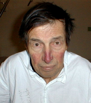
General- May see obvious resp distress, mitral facies (i.e. pink-purple patches from vasoconstriction on face)
Pulse: May see A-Fib. Usually normal, but may see low volume.
JVP: May see signs of pulmonary HTN as consequence of MS
Palpation: may have tapping S1 (pliable anterior leaflet). In LLD, may palpate diastolic thrill. May have RV heave and palpable P2 from pulm HTN.

Heart sounds:
Major findings are
1) loud S1 early, soft late in course
2) opening snap- early diastolic sound of MV opening; moves closer to S2 with more severe
3) diastolic rumble (murmur) post- opening snap
4) late diastolic murmur with presystolic accentuation from atrial kick


Others:
1) ECG- L atrial enlargement- 'p-mitrale'
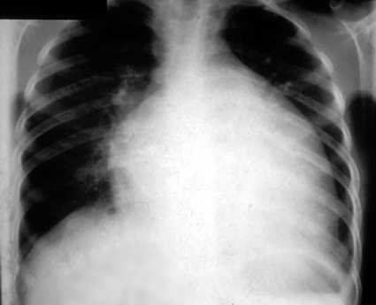
2) CXR- splayed carina, double R heart border, flat or everted L heart border from LA enlargement


Link:
Click here for a nice website from the Cleveland Clinic summarizing MS

Monday, March 15, 2010

Thrombocytopenia and ITP


Today we discussed an approach to a bleeding diathesis, and issues related to thrombocytopenia and ITP. A few key points:

General approach to bleeding history:
Try to differentiate primary hemostasis problem (vascular response and platelet plug) vs. secondary hemostasis problem (fibrin clot, or resolution phases)

Clues:
Primary- excessive immediate post-traumatic bleeding usually. Petechiae, purpura, mucosal bleeds: nose, mouth, gums, GI, uterine
Secondary- delayed, subcutaneous, deep tissues, joints, GI/GU, post-op.
Acquired vs. inherited: new-onset vs. lifelong, new medical problem or drug, FHx.


General approach to thrombocytopenia

3 major categories
1. Decreased platelet production
2. Increased platelet destruction or platelet consumption
3. Sequestration

Decreased PLT production: Bone marrow problem; marrow shows decreased megakaryocytes
Aplastic anemias (congenital or acquired)
Myelophthisic anemia - i.e. marrow replacement (malignancy, granulomatous, etc)
Megaloblastic anemias (B12, folate deficiency)
Marrow suppression from drugs, toxins, infection, etc)

Peripheral destruction or consumption
Immune-mediated: ITP- Primary (idiopathic) or secondary (SLE, HIV, EBV B-cell disorders), drug-induced, post-transfusion
Non-immune: Thrombotic thrombocytopenia purpura- TTP, disseminated intravascular clotting

Sequestration:
Usually portal hypertension, hypersplenism

ITP specifics

Most cases are idiopathic.
In adults, it is generally chronic, the onset is often insidious. Approximately
twice as many women as men are affected

If all these conditions are met, bone marrow bx (gold standard) is not required
-age less than 60
-isolated TCP
-no splenomegaly
-no lymphadenopathy

To exclude before calling "idiopathic ITP":
-non-immune causes of TCP (primary marrow problem, TTP, HIT, DIC, etc)
-infection- HIV especially, EBV
-inflammatory disorders, esp. SLE
-malignancy, esp. B-cell disorders (CLL, myeloma, etc)
-drug induced (for immune-mediated, B-lactams, quinine, vancomycin, others)

Therapy:
In serious bleeding
1) hemodynamic and RBC transfusion support
2) platelet transfusion
3) steroid
4) IVIG
5) some evidence for factor VII in life-threatening bleeding

Most pts require therapy; those who do not are those with mild (over around 50), asymptomatic, incidentally discovered thrombocytopenia. Otherwise, options are:
-Steroids (prednisone 1mg/kg, tapered over 4-6 weeks depending on response or dexamethasone 40mg PO x 4d). 50% have relapse. RCT underway comparing these regimens
-IVIG; indicated for severe bleeding, pre-operatively or pre-delivery, and in steroid-refractory cases
-Splenectomy (need appropriate vaccinations first)
-Rituximab (controversial whether should be tried before splenectomy)

In chronic refractory cases, there may be a role for vincristine, azathioprine and cyclophosphamide, and thrombopoeitin agonists

Links:

Click here for a NEJM review of ITP
Click here for a series demonstrating effect of short course of dexamethasone described above

Thursday, March 11, 2010

Hypocalcemia


Today we discussed hypocalcemia. A few points:

Clinical manifestations:
-Paresthesias
-Lethargy
-Seizures
-possibly arrhythmias (lengthens QT)

-Chvostek's sign:
Tap cheek 2cm anterior to tragus, look for ipsilateral upper lip twitch
NB- this video is freely available on YouTube, and is not a patient cared for in this hospital

-Trousseau's sign:
Inflate BP cuff above systolic pressure for 3 minutes, look for painful carpal spasm (shown above)

An approach to causes of hypocalcemia (which drives appropriate history, physical, and investigations):

Think of major determinants of calcium handling, and what can affect each:

Low PTH:
-iatrogenic (most common cause); e.g. post-thyroidectomy
-autoimmune (can be part of autoimmune polyendocrine syndrome with vitiligo and candidiasis)
-infiltration of the parathyroids (sarcoid, hemochromatosis)
-hypomagnesemia (alters PTH receptor sensitivity)

Low Vitamin D:
-malabsorption of fat-soluble vitamins (ADEK)
-liver disease
-kidney disease
-inadequate sun exposure

GI tract:
-malabsorption of calcium (proximal small bowel): celiac, Crohn's, others in addition to vit D malabsorption

Kidneys:
-Loop diuretics
-Calcium wasting: Fanconi's syndrome (proximal tubular reabsorption problem; causes hypocalcemia, hypophosphatemia, non-anion gap metabolic acidosis, hypoglycemia, hypoalbuminemia)

Bone:
-"Hungry bone syndrome"- post parathyroidectomy after hyperparathyroidism, increased bone uptake of calcium after prolonged calcium "leaching"
-osteoblastic metastases (esp. prostate, breast)

Intravascular Ca binding:
-citrate (e.g. in dialysis or massive transfusion)- binds ca
-hyperphosphatemia

Calcium deposition sites:
-severe pancreatitis
-rhabdomyolysis

Others:
-secondary hyperparathyroidism; often have normal or low Ca in renal failure for many reasons
-DiGeorge syndrome- affects PTH receptor
-Bartter's syndrome- acts like a loop diuretic causing calciuresis
-"pseudohypoparathyroidism"- looks like hypoparathyrodism, except PTH is high; receptor mutation

Tip: The phosphate level can be quite helpful in sorting out causes; PTH increases calcium and decreases phosphate. Vitamin D increases both calcium and phosphate.

Link:
Click here for a NEJM review of hypoparathyroidism that discusses much of above

Wednesday, March 10, 2010

Peripheral neuropathy


Today we discussed peripheral neuropathy. A few points on this very broad topic:

Important differential: 'THIN'
1) Toxic, Metabolic- medications, DM2, EtOH, metals, B12
2) Hereditary- Charcot-Marie-Tooth and variants
3) Inflammatory and infectious- Guillain-Barre, CIDP (chronic inflammatory demyelinating polyneuropathy), vasculitis, and HIV, HTLV, leprosy
4) Neoplastic/paraneoplastic: multiple myeloma

Important points on history:
1) Temporal course- acute (days to weeks), subacute (4-8 weeks), chronic (>8 weeks); relapsing? progressive?
2) Proximal vs. distal motor and sensory symptoms (trouble reaching overhead, climbing stairs vs. opening jars, picking things up)
3) What are the sensory symptoms? negative- e.g. numbness or positive- e.g. paresthesias, burning, throbbing, etc.
4) Questions around above differential

Important physical exam:
Atrophy, fasiculations for LMN lesion
Distribution of weakness- nerve vs. plexus vs. root
Reflexes
Sensory- pin prick, vibration (named nerve vs. nerve root vs. length-dependent)

Important investigations:
FBG, HbA1C
CBC, Cr, Vitamin B12, TSH, serum and urine electrophoresis, HIV

Multiple named nerve palsies = mononeuritis multiplex
Essentially diabetes-related vs. vasculitis.
Common nerves involved in DM2: CN III, IV, VI, median nerve, peroneal nerve.
Vasculitides that commonly cause mononeuritis multiplex: Churg-Strauss, polyarteritis nodosa, rheumatoid arthritis with vasculitic component

Link:
Click here for an excellent review of peripheral neuropathy from Lancet

Tuesday, March 9, 2010

Pancreatitis


Indeed...


Today we discussed an approach to pancreatitis. A few points:

Acute = no pre-existing pancreatic pathology; usually resolves without permanent damage.
Chronic = pre-existing fibrosis/scarring/calcification. EtOH-induced is often acute on chronic.

Pancreatitis is essentially autodigestion, release of zymogens, and in severe cases, a retroperitoneal chemical burn injury.

Etiologies:
-EtOH
-Gallstone
-Drugs (commonly reported include some HIV medications, HRT, azathioprine, tetracycline, 5'ASA, sulfasalazine)
-Metabolic- hypertriglyceridemia, hypercalcemia
-Iatrogenic (esp. post-ERCP)
-Autoimmune
-Others- anatomic (pancreas divisum), scorpion bites (everyone's favourite), cystic fibrosis, infection

Symptoms: Epigastric abdo pain, better leaning forward (Ingelfinger's sign for medical jeopardy fans), radiating to back, N+V

Things to look for on exam:
Vitals: Tachypnea, tachycardia, fever
-jaundice
-abdominal distension, absent bowel sounds (ileus)
-cullen/grey turner signs
-dull lung bases from pleural effusion
-abdominal tenderness.

Lab: Lipase/amylase (NB- macroamylasemia, salivary gland pathology, and tubo-ovarian pathology give high amylase but normal lipase). Lipase and amylase support the dx, not make it. The levels fall off in days.

Imaging:
AXR: may show colon-cutoff sign or ileus
CT may show pseudocyst or necrosis in addition to stranding, inflammation

Prognostic factors: Development of organ failure, SIRS (look for ARF-most common, ARDS), necrosis, abscess, pseudocyst. Others: Ranson criteria, APACHE score, etc.

Therapy:
Largely supportive; monitor closely for complications.
Principles of managment:
1) Fluid resuscitate aggressively as needed
2) Watch respiratory status, and intervene if necessary (ABG's, etc)
3) Watch for other organ involvement (AKI, DIC, etc)
4) Nutrition: Do not rest the pancreas; start enteral nutrition (increased metabolic requirements, decreased bacterial translocation) - evidence for early PO feeding.
5) Pain managment
6) Antibiotics- very controversial; only convincing evidence is in pancreatitis with necrosis (there are scoring systems based on CT), in which case meropenem has proven benefit. In some cases, a fine needle aspirate of a collection is done to determine whether infected and only treated if positive.
7) If gallstone-related, ERCP and cholecystectomy (during current admission if mild or delayed if severe) reduces recurrence risk from 25% to 8%.

If severe deterioration, consider surgery for debridement, but this is high-risk.

Links:
Click here for an interesting paper highlighting common problems and pitfalls in pancreatitis management
Click here for a Cochrane review of antibiotics in pancreatitis
Click here for a trial comparing symtom-drive vs. standing benzos for alcohol withdrawal