Tuesday, July 19, 2011
Infective endocarditis
Morning report today was a case of presumed endocarditis in a VERY susceptible host. See a prior post here with a link at the bottom to the AHA 2008 update.

ECG limb-lead reversal
 Amuse-bouche yesterday was an ECG with limb-lead reversal. These are important to pick-up as some can mimic clinical scenarios.
Findings and clinical mimics of common limb-lead changes:
Right arm-Left arm:
- Q wave I, aVL
- Inverted p-wave I, II, II, aVF
- Mimics: old lateral infarct, non-sinus atrial activity, dextrocardia (limb lead reversal will still have normal R-wave progression, dextrocardia will not)
Right arm- Left leg:
- Q wave II, III, aVF
- Inverted p-wave II, III, aVF
- Mimics: old inferior infarct, non-sinus atrial activity
Right arm- Right leg:
- Diffuse low voltage in limb leads (esp. lead II)
- Mimics: any condition causing low voltage (limb-lead reversal will have normal precordial voltages)
Other limb lead reversals causing only minor changes with no real clinical mimics include Left arm- Left leg, Left arm- Right leg and leg-leg reversals. Precordial lead changes will interfere mostly with R-wave progression.
More from an old but concise review here.
Monday, July 18, 2011
Thrombotic thrombocytopenic purpura
Morning report today was a great case of Thrombotic thrombocytopenic purpura (TTP). Please see here for a prior post on TTP, with links at the bottom to two good NEJM articles.
Friday, July 15, 2011
Mesothelioma
 AB today was on mesothelioma.
A few points on mesothelioma, with more in NEJM here:
–Malignancy of pleura (<90%) or peritoneum (>10%)
–~90% of patients will have a history of exposure to asbestos, with mean time from exposure to diagnosis ~35y (wide range)
-Usually presents with local pain, cough or dyspnea, and often assocaited with weight loss/fatigue
- Pleural biopsy (VATS or open) is usually definitive, but special stains are required to differentiate from adenocarcinoma
- Despite some advances, including agressive multi-modal therapy (chemo/radical surgery/radiation) and new active agents (pemetrexed), survival is poor with <10% surviving past 2 years.
- Although Canadian and US incidence is dropping (likely due to asbestos regulation), the overall worldwide incidence is increasing- for which Canada is playing no small part (see here).
Hyponatremia
Morning Report today was on hyponatremia. See an old post here for a good overview. There are two good papers linked at the bottom of the post.
Thursday, July 14, 2011
Familial Adenomatous Polyposis
 Amuse-bouche today was Familial Adenomatous Polyposis (FAP). This results from a mutation of the tumor suppressor gene APC on chromosome 5. A quick review of the features:
- Upper-GI tumors: Fundic gland polyps, duodenal adenomas
- Adenocarcinoma of the ampula of Vater
- Extra-intestinal features: osteomas, desmoid/soft-tissue tumors, retinal pigment hypertrophy
Here is a link to a basic BMJ review on hereditary colorectal cancer.
Renal failure in multiple myeloma
Today in morning report, a good case of renal failure in multiple myeloma. A good prior post on all of the causes here with a link at the bottom to a review article.
Wednesday, July 13, 2011
Dermatomyositis
Amuse-bouche today was Gottron's papules and dermatomyositis. A prior post on dermatomyositis here.
A quick review of the associated dermatologic findings:
Gottron’s papules-80% of patients
Hyperkeratotic, erythematous, flat papules
Dorsum MCP and IP joints, less commonly wrists/elbows/knees
Heliotrope rash- <50% of patients
Periorbital violaceous/erythematous rash
One or both eyelids
May be accompanied by edema
Shawl sign
Macular erythema in V shape at nape of neck
Mechanic's hands
Rough and scaly with fissuring
Lateral and palmar areas of fingers
Nail changes
Periungal erythmea
Telangiectasias on the proximal nail fold
A quick review of the associated dermatologic findings:
Gottron’s papules-80% of patients
Heliotrope rash- <50% of patients
Shawl sign
Mechanic's hands
Nail changes
Renal failure and hyperkalemia
In morning report today was a case of acute renal failure. A good prior post with a link at the bottom to a good NEJM article here.
We didn't get in to the management of hyperkalemia, but as Dr. Bunce said- you should know it by tomorrow, so here is a prior post with a good link to a review at the bottom.
We didn't get in to the management of hyperkalemia, but as Dr. Bunce said- you should know it by tomorrow, so here is a prior post with a good link to a review at the bottom.
Tuesday, July 12, 2011
Pendred's syndrome
The amuse-bouche today was Pendred's syndrome. A link to a good article here.
Pendred's syndrome is an autosomal-recessive disorder of iodine organification caused by mutations in iodine transport protein pendrin.
It is characterized by the combination of congenital sensorineural hearing and goiter, and accounts for up to 10% of cases of hereditary deafness.
Patients have a positive perchlorate discharge test because they cannot provide substrate for the organification step in thyroid hormone synthesis.
Pendred's syndrome is an autosomal-recessive disorder of iodine organification caused by mutations in iodine transport protein pendrin.
It is characterized by the combination of congenital sensorineural hearing and goiter, and accounts for up to 10% of cases of hereditary deafness.
Patients have a positive perchlorate discharge test because they cannot provide substrate for the organification step in thyroid hormone synthesis.
Decompensated cirrhosis

Morning report today was a case of fever and decompensated cirrhosis. A prior post on cirrhosis with a link to a good lancet article on management here.
A reminder of the Child-Pugh score above, with total points giving grades A (5-6 ) B (7-9) and C (10-15) and higher mortality with inreasing points/grade.
Monday, July 11, 2011
Seizures and alcohol withdrawl
Plasmacytoma and multiple myeloma
Thursday, July 7, 2011
CLL and stem cell transplants
 Today in morning report, and interesting case of fever in CLL. Here is a link to a prior post on CLL and immunodeficiency; see the bottom of the post for a link to the role of IVIG: http://morningreporttgh.blogspot.com/2010/04/cll-and-its-complications.html
The other discussion was on stem cell transplants and graft versus host disease (GVHD). A few points:
1) Types of stem cell transplant:
Autologous: Patient's own mononuclear cells are harvested from bone marrow or (now more commonly) from peripheral blood prior to high-dose myeloablative chemotherapy/radiotherapy. The cells are then reinfused during the period of prolonged bone marrow failure that occurs after these treatments in order to help restore hematopoiesis. Classically this type of transplant was used in lymphoma, but there is an ever-expanding list of trials for other indications including solid tumors and plasma-cell dyscrasias.
Allogeneic: Stem cells are harvested from the bone marrow of healthy donors or newborn umbilical cord/placenta. The cells are infused to reconstitute the patient's hematopoeitic system, but also can have direct anti-leukemia activity, termed graft-versus-leukemia (GVL). Given that the cells are from a donor, there are a greater number of risks than in autologous transplant due to immune suppression and graft-versus-host-disease (below).
2) Graft versus host disease:
Lymphocytes from the donor may target normal host tissues and cause inflammation, with particular predilection for skin, gut, liver, and lungs. The acute form usually occurs before 100 days post-transplant and can present with a maculopapular or bullous rash, cholestasis potentially leading to hepatic failure, or secretory diarrhea. The chronic form more resembles other autoimmune disorders, presenting with arthritis, scleroderma-like skin changes, sicca syndrome, chronic hepatitis, and GI malabsorbtion.
The risk of GVHD is directly related to age and inversely related to the the degree of major histocompatibility antigen matching between donor and recipient. It remains the major complication of allogeneic transplant, affecting upwards of 40% of patients.
Wednesday, July 6, 2011
Brugada syndrome
The answer to this morning's amuse-bouche was Brugada syndrome. More from an old blog post here: http://morningreporttgh.blogspot.com/2010/03/brugada-syndrome-and-arrhythmogenic-rv.html
Pericardial effusion
Morning report today was on pericardial effusions.
Here is a prior post on the causes of pericarditis
http://morningreporttgh.blogspot.com/2009/04/shot-through-heart-and-your-to-blameyou.html.
When thinking of pericardial effusions, the causes are all the same, as well as the addition of hypothyroidism.
We talked only briefly about cardiac tamponade, but here is a link to the JAMA rational clinical exam paper 'Does this patient have cardiac tamponade'
http://jama.ama-assn.org/content/297/16/1810.abstract
Here is a prior post on the causes of pericarditis
http://morningreporttgh.blogspot.com/2009/04/shot-through-heart-and-your-to-blameyou.html.
When thinking of pericardial effusions, the causes are all the same, as well as the addition of hypothyroidism.
We talked only briefly about cardiac tamponade, but here is a link to the JAMA rational clinical exam paper 'Does this patient have cardiac tamponade'
http://jama.ama-assn.org/content/297/16/1810.abstract
Blogging is so 2010
 It's early July: the season of bad tan lines and new chief medical residents (in this case maybe both in one package...just you wait!)
I will be using the blog quite a bit this year, but I plan to use it a little differently than before in a few ways:
(1) I will try to post daily:
I want to spare your email inboxes, so I will post daily on this site, and you can check things out as you see fit for your own learning goals.
(2) I will not really be blogging:
The posts will usually be shorter, less like a true blog and more similar to twitter-style tweets, with links to papers or other resources (including prior blog posts from TGH, TWH, and MSH morning report blogs).
However, I will write longer posts on topics that haven't been covered before and can be reasonably covered in without too much text .
(3) I will also tweet:
For those of you who already have twitter or are interested in using it, I will tweet the same links as I blog @tghcmr (http://twitter.com/#!/search/tghcmr)
I am here to help you learn internal medicine this year, so if you find none of this works well for you, let me know, and I can always make changes.
Cheers,
Chris
Wednesday, November 24, 2010
Renal Tubular Acidosis and Fanconi Syndrome

Today is my last post here on Horses and Zebras. Thanks for the good times. The banner will be picked up again by the next CMR come January.
This morning we discussed Fanconi Syndrome and touched briefly on Renal Tubular Acidosis (RTA).
Here are two links to review articles, one from the Journal of the American Society of Nephrology and one from the Archives of Internal Medicine.
Some points about RTA:
1) In the setting of an acidosis, the kidneys maintain acid/base homeostasis by increasing resorption of HCO3 and increasing H+ excretion. HCO3 resorption occurs predominantly in the proximal convoluted tubule (85-95%) with the remainder occurring in the distal convoluted tubule (10%). The secretion of H+ typically occurs in the DCT as ammonium.
2) In RTA, the kidney loses the ability to perform one of these functions and a non-anion gap (hyperchloremic) metabolic acidosis develops.
3) There are 3 types of RTA:
a) Type 1 (distal)
b) Type 2 (proximal)
c) Type 4 (hypoaldosteronism)
(Type 3 RTA is a mix between types 1 and 2 and is seen in a rare genetic disorder)
4) Type 1 (distal) RTA:
a) Results in decreased ammonium secretion in the DCT causing systemic acidosis (HCO3 less than 10).
b) Most common adult causes include: autoimmune diseases (Sjogren's, RA, SLE), hyperglobulinemia, meds (lithium, amphotericin B), hypercalciuria, liver disease and sickle cell disease.
c) Often presents with NAG metabolic acidosis, hypokalemia, hypercalciuria and an elevated urine pH (greater than 5.5).
5) Type 2 (proximal) RTA:
a) Can occur in isolation, but is most commonly seen in the setting of diffuse proximal tubular dysfunction (Fanconi syndrome - as discussed today).
b) The typical findings in Fanconi syndrome include: bicarbonaturia, glucosuria, phosphaturia, uricosuria, aminoaciduria and tubular range proteinuria.
c) Isolated Type 2 RTA presents with bicarbonaturia due to impairment in bicarbonate resorption in the PCT.
d) While the bulk of HCO3 resorption occurs in the proximal tubule, some occurs in the DCT. In Type II RTA, there is a threshold level of serum HCO3 above which filtered the HCO3 overwhelms the resorptive capacity of the DCT and HCO3 loss in the urine occurs. The borderline is generally around 16mmol/L.
e) The most common cause of Fanconi syndrome in adults is multiple myeloma (due to light chains), acetazolamide use and some chemotherapeutic agents.
6) Type 4 (hypoaldosteronism) RTA:
a) Characterized by a deficiency in, or tubular resistance to, the action of aldosterone.
b) Typically presents with hyperkalemia and a mild acidosis.
c) Most common causes include: diabetic nephropathy, CKD, primary adrenal pathologies and medications that inhibit the RAAS.
7) Differentiating Type I from Type II:
a) Consider calculating the urine anion gap (Na + K - Cl) on a random sample. The Cl- is an indirect marker for NH4+ secretion and thus a positive value supports a dx of Type I RTA (reduced H+ secretion) whereas a negative value may suggest Type II.
b) An increased urine osmole gap can also suggest Type I RTA.
c) The metabolic effects of Fanconi syndrome are only found in Type II RTA.
d) Urine pH will always be elevated in Type I RTA but will be variable and increase in Type II RTA if the serum HCO3 is raised beyond the reabsorbing threshold of the DCT (since the PCT is dysfunctional).
8) Management of Type I and II RTA typically involves reversing any causative conditions and providing oral alkali either as sodium bicarbonate or citrate. Type IV RTA is managed again by addressing the underlying etiology and providing aldosterone replacement in the form of fludrocortisone.
Tuesday, November 23, 2010
Streptococcus Anginosus Empyema
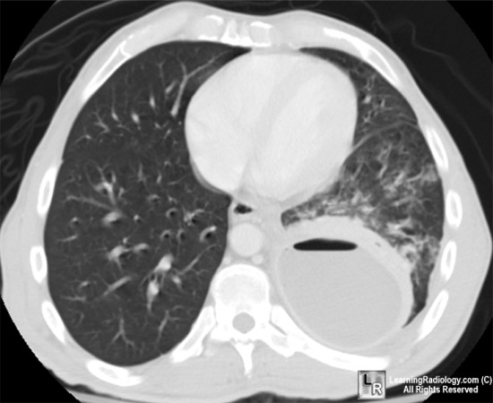 Today we discussed a patient with a chronic empyema ultimately proven to be secondary to streptococcus anginosus.
Please follow the link to a recent review on parapneumonic effusions and empyema.
Some take-home points about S.anginosus and empyema:
1) The Streptococcus milleri group of organisms is made up of S. intermedius, S. anginosus and S. constellatus which are considered a sub-group of Viridans streptococci. These gram positive cocci are normal flora in the respiratory and GI tract and are known to cause abscesses in the brain, liver, lungs and oropharynx.
2) S.milleri are generally susceptible to beta-lactam antibiotics.
3) In general, lung abscesses related to S. milleri are thought to occur from aspiration of oropharyngeal contents.
4) Don't forget to look for the caramel of butterscotch smell associated with S. anginosus infections! Actually no, please don't.
5) The management includes prompt drainage of any collections and IV antibiotic therapy, usually with a beta-lactam. Duration of treatment is dependent on symptoms, but often a minimum 4 week duration of IV antibiotics is required. Specific antibiotic selection should be based on sensitivity testing.
6) Regarding complicated parapneumonic effusions and empyema:
a) These effusions typically occur in the context of pneumonia and represent a spectrum from an uncomplicated parapneumonic (UPPE) effusion to a complicated parapneumonic effusion (CPPE)to an empyema.
b) The signs and symptoms of an empyema are often indistinguishable from a typical pneumonia. All patients with pneumonia should be assessed for a pleural effusion.
c) In the setting of pneumonia with a pleural effusion, the phrase "never let the sun set on a parapneumonic effusion" is used to emphasize the importance of prompt investigation and drainage if necessary. Effusions greater than 10mm on the lateral decubitus CXR should be sampled.
d) More chronic presentations of an empyema can occur with less virulent organisms such as S.anginosus (as in our patient).
e) In general, a PPE should be drained if the pH of the fluid is less than 7.2, there is a positive gram stain/culture, a glucose less than 40-60 (U.S. units), an LDH greater than 1000 (U.S. units) or the effusion is greater than 50% of the volume of the hemi-thorax.
f) Drainage of a CPPE or empyema typically involves a large bore chest tube +/- adjunctive therapies such as fibrinolytics, VATS or decortication.
Monday, November 22, 2010
Antibiotics and Pneumonia

Today we talked today about a patient with community acquired pneumonia (CAP) and the appropriate antibiotics and investigations for CAP.
Here's a link to the 2007 IDSA/ATS CAP Guidelines (scroll down to CAP). The BTS also released guidelines in 2009.
In brief:
1) While it seems obvious, an infiltrate on CXR (in the right clinical context) is required for the diagnosis of pneumonia.
2) Generally, all patients with pneumonia admitted to hospital should have blood and sputum cultures drawn.
3) In patients with severe pneumonia, urinary antigens for Legionella pneumophila and Streptococcus pneumoniae should be sent.
4) Options for outpatient treatment:
a) If previously healthy, can use a macrolide or doxycyline.
b) In patients with comorbidities or recent Abx use, give a respiratory FQ or a (beta- lactam + a macrolide).
c) Remember, if the presence of macrolide resistant S.pneumo is greater than 25% in your practice area, macrolides should be avoided.
5) Inpatient, non-ICU treatment:
a) (Respiratory FQ) or a (beta-lactam + a macrolide).
6) Inpatient, ICU treatment:
a) A beta-lactam plus either a macrolide or a FQ (better evidence for FQ over macrolide).
b) If concerned re: pseudomonas, give an anti-pseudomonal beta-lactam that has anti-pneumococcal activity (PipTazo, Cefepime, Imipenem or Meropenem) plus either (Cipro of Levo) or (an aminoglycoside + azithromycin) or (an aminoglycoside + an anti-pseudomonal FQ).
c) Add Vano or Linezolid if MRSA risk factors present.
7) The beta-lactam in the outpatient and in-patient not ICU treatment arms can be high dose amoxicillin/ampicillin.
Subscribe to:
Posts (Atom)